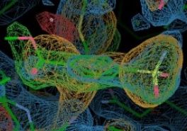
Macromolecular Crystallography (MX)
Access to Diamond MX beamlines is now operating under these guidelines. We are able to offer remote access, unattended automated data collection and mail-in services for all research areas as well as on site user access.
Access to unattended automated data collection is available to all users.
Diamond provides a range of techniques for academic and industrial researchers studying the machines of life. As one of those techniques, Macromolecular Crystallography (MX) reveals the shape and arrangement of biological molecules at atomic resolution, knowledge of which provides a highly accurate insight into function. This can be combined with complementary information from many other techniques available at Diamond alongside lab based investigations to reveal the broader picture of molecular interactions and their effects.
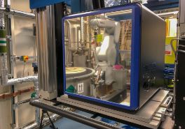
MX is a core activity at Diamond with seven beamlines dedicated to the technique alongside the XFEL Hub, Membrane Protein Laboratory and XChem fragment screening facility for the extensive UK structural biology community as well as researchers in Europe and beyond. The staff of the MX group are recognised as innovative world leaders in MX, moving the goalposts of what is feasible for 'conventional' MX as well as developing techniques and beamlines that transform MX to the next level, enabling new experiments and methodologies. The group takes a long term approach to enabling new capabilities at its suite of beamlines to meet the current and future demands of an exacting community of scientists.
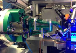
Microfocus
Specialised high flux microfocus beams for microcrystal data collection or the illumination of subvolumes of larger inhomogeneous crystals.
More information...Remote Access
The highly automated beamlines enable data collection from anywhere in the world via remote access. Systems are in place for shipping samples to and from Diamond Light Source. New users can borrow pucks and tools.
More information...In situ Data Collection
In situ data collection and sample characterization directly from crystals in their crystallization plates.
More information...Sample Humidity Control
Control of the sample crystal's relative humidity with the aim of improving diffraction quality through improved packing of protein molecules constituting the lattice.
More information...Spectroscopy
On-line and off-line UV-Vis spectroscopy of macromolecular samples to study enzyme reaction mechanisms and radiation damage.
More information...Multi-axis Goniometry
Crystal reorientation to optimize data collection strategies for anomalous diffraction data collection and low symmetry spacegroups, or very long axis unit cells.
More information...Biocontainment
All beamlines are designated bio containment level 1. For containment level 2 samples beamlines I03 and I24 can be used and beamline I03 can be configured to run at biocontainment level 3.
More information...Fragment Screening - XChem
The fragment screening facility at Diamond Light Source is based in Lab 36 and on beamline I04-1. The scope of the facility is to encompass all stages of the fragment screening experiment.
More information...VMXm
Commissioning
VMXm is a micro/nanofocus MX beamline aimed at atomic structure determination in cases where the production of significant quantities of protein material and crystals is problematic.
More informationDetector: Pilatus3 6M (Si) and Eiger2 X 9M (CdTe)
Wavelength: 0.44 - 1.77 Å
Energy: 7.0 - 28.0 keV
VMXi
Operational in optimisation mode
The Versatile Macromolecular X-tallography in-situ (VMXi) beamline will be an entirely automated facility for characterisation of, and data collection directly from, crystallisation experiments in situ.
More informationEnergy: 16 keV
I03 MX
High throughput and fully automated beamline for optimised MX experiments. Capable of accepting CL3 type experiments on crystals of pathogens.
More informationDetector: Eiger2 XE 16M
Wavelength: 0.50 - 2.07 Å
Energy: 6 - 25 keV
I04 Microfocus MX
Variable focus from 5 to 100 microns, high throughput and highly automated beamline for optimised MAD and SAD experiments.
More informationDetector: Eiger2 XE 16M
Wavelength: 0.69 - 2.07 Å
I04-1 Fixed wavelength MX
High throughput and highly automated fixed wavelength SAD beamline for macromolecular crystallography.
More informationDetector: Eiger2 XE 9M
Wavelength: 0.92Å (fixed)
Energy: 13.445 keV
I23 Long-Wavelength MX
Operational in optimisation mode
A unique facility for solving the crystallographic phase problem, using the small anomalous signals from sulphur or phosphorous which are present in native protein or RNA/DNA crystals. Additionally, anomalous difference Fourier maps can be used to locate sulphur and phosphorous positions to assist model building at low resolution and/or identify lighter atoms such as chlorine, potassium and calcium.
More informationEnergy: 2.1 – 11 keV
I24 Microfocus MX
High throughput variable microfocus beamline for optimised MAD and SAD experiments on crystals down a few microns in size.
More informationDetector: Pilatus3 6M
Wavelength: 0.62Å - 1.77Å
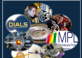
Membrane Protein Laboratory
The Membrane Protein Lab (MPL) at Diamond is a research and training facility for scientists interested in solving the 3D structures of membrane proteins by X-ray crystallography.
More informationXFEL Hub
The UK is taking a leading role in the development of a new structural biology facility (SFX) at the European X-ray Free Electron Laser (XFEL), in Hamburg, Germany, and a complementary facility at Diamond (The UK XFEL Hub) to help develop the required expertise.
More informationXChem - Fragment Screening
The fragment screening facility at Diamond Light Source is based in Lab 36 and on beamline I04-1. The scope of the facility is to encompass all stages of the fragment screening experiment.
More informationCrystallisation Facility
The Crystallisation Facility @ Harwell is a research and training facility for the scientific community interested in solving the 3D structures of soluble or membrane proteins by X-ray crystallography.
More information
| Beamline | I03 | I04 | I04-1 | I23 | I24 |
| Wavelength range (Å) | 0.6 - 2.3 | 0.69 - 2.07 | 0.92 (fixed) | 1.18 - 5.9 | 0.7 - 2.0 |
| Energy range (keV) | 5.2 - 21.0 | 6.0 - 18.0 | 13.5 (fixed) | 2.1 - 11.0 | 6.4 - 20.0 |
| Default settings (Å/keV) | 0.98 / 12.7 | 0.98 / 12.66 | 0.92 / 13.53 | 2.755 / 4.5 | 0.97 / 12.8 |
| Flux (ph/s) in full beam at default energy at 300 mA | 1.0 x 10¹³ | 2.0 x 10¹² | 3.8 x 10¹² |
7.5 x 10¹⁰ @ 100 x 100um |
3.0 x 10¹² |
| Default beamsize (µm) | 90 x 30 | 30 x 20 | 60 x 50 | 100 x 100 | 8 x 8 |
| Full beam size at sample (µm) | 90 x 20, 90 x 30, 100 x 100, 55 x 10 | 10 x 5 --> 110 x 100 | 60 x 50 | Any (slits) |
5 x 5 --> 50 x 40 |
| Available apertures (µm) |
20, 50, 100 |
10, 20, 30, 50 and 70 | (50 - 600) x (50 - 600) | ||
| Detectors (Dectris type) |
Eiger2 XE 16M (560 Hz) |
Eiger2 XE 16M (560 Hz) |
Eiger2 XE 9M (560 Hz) |
Custom Semi-cylindrical P12M (10Hz) |
P3-6M (100 Hz) EIGER2 X CdTe 9M |
| Multi-Axis Goniometry | Yes | Yes | No | Yes | Yes |
| Number of unipucks / SPINE pins | 37/592 | 37 / 592 | 37 / 592 | 20 I23 specific pins | 37 / 592 |
| Maximum samples / hour | 30 | 30 | 30 | 6 | 30 |
| Typical samples / hour | 15 - 25 | 15 - 25 | 15 - 35 | 1 - 6 | 15 - 25 |
| Minimum Detector Distance (mm) | 140 | 170 | 159 | N/A (0) | 200 |
Plan
Reserve access well ahead of your visit either at the links in the web page or via your local contact to facilities such as
Check the Diamond MX web pages and individual beamline pages for updates, manuals and contact details prior to your experiment.
Register your shipping dewars with Diamond to use our shipping account and help with tracking.
Always purchase and use SPINE pins and Unipucks to mount and ship your samples. Non-SPINE standard pins cause robots to crash and damage to equipment: you lose beamtime, we lose sleep.
For full remote access experiments we prefer pucks to arrive in the shelved shipping cane or alternatively the puck shipping canister. Please avoid the narrow canisters where possible.
Plan
For remote experimenters read the remote access guidelines.
- For remote access experiments the team leader should update UAS with contact details for all remote users, and ensure all pucks are correctly assigned on ISPyB at least two working days before the visit.
- For mixed remote access (i.e. where a member of your team is on-site) ensure they will be available to change pucks if required.
- In both cases discuss your requirements for remote access with your local contact before your experiment.
For all experiments we highly recommend preparing your shipments in advance in ISPyB, identifying your pucks by the puck identifier (e.g. CPS-0024) and assigning samples at the same time. This allows pucks to be assigned to robot positions as loaded by either you or Diamond staff making them available in GDA immediately. For remote access this is a requirement rather than a recommendation to ensure reliable information transfer.
For UK users with Diamond registered dewars you can book your next working day shipping to us with Diamond’s DHL account. We recommend shipping your dewars three or more days before your experiment to ensure arrival in time.
Register your PDB and sequences for your proteins in ISPyB. These can then be used automatically if you use default file names in GDA for all future visits in downstream processing such as XIA2, DIMPLE and MrBUMP and avoids the need to do this for each visit.
Refer to the manual for data collection possibilities –
- grid scans - find your crystals or the best diffracting region of a crystal and centre reliably
- line scans – minimise radiation damage, maximise resolution
- fluorescence data – find out if you have metals and what energy to use to exploit for phasing
- in-situ experiments - test crystal and collect data in trays
- dehumidifier HC1 - optimise diffraction by adjusting humidity
- data collections – optimise your data collections:
- strategies
- multi axis goniometry
- inverse beam
- interleaved MAD
- washing and annealing crystals and other tools.
Read the message of the day in GDA for beamline status.
Refer to beamline specific manuals where appropriate.
Start your data back-up as soon as you can.
Load your pre-registered pucks and assign positions on the touch screen in the hutch (soon in all beamlines). Now start GDA and all your samples will be preloaded in the sample changer view (or press refresh to update if GDA was already running).
Use default file names in GDA to streamline experiments, avoiding the need to type information at each sample load, preventing mistakes and ensuring downstream processing uses the information you have provided in advance (PDB, sequence, space group).
Monitor your results from our automatic software pipelines in ISPyB.
Talk with your local contact about your experiment plans – we are here to help you solve structures.
Unload your last sample with the sample changer.
If on site, with sufficient time to ensure you have removed pucks from the sample changers before the end of your shift.
If using data backup via disk; Finalise your backup disk and unmount cleanly in the data dispenser interface.
Else: Transfer required data using globus.
See Shipping Dewar Home, for details on arranging return shipment
Diamond pays for sample shipping (2 dewars per shift) for EU and UK users, if using our shipping agent DHL via ISPyB. No other shipping method can be reimbursed.
You can reprocess your data afterwards on our cluster.