• Diamond Home page
  • About
  • For Users
  • Public
  • Industry
  • Instruments
  • Careers
  • More Show more menu items
Search

About

  • About Diamond
  • About Synchrotrons
  • News and Features
  • Events
  • Contact Us
  • FAQ

For Users

  • Apply for beamtime
  • User guide
  • Diamond Users Commitee
  • FAQ
  • User Administration System
  • iSpyB

Industry

  • Techniques Available
  • Industry Research
  • Industry Case Studies
  • News
  • Meet the Industry Team

Science

  • Research
  • Computing
  • The Machine
  • Publications
  • Research Expertise
  • Membrane Protein Laboratory
  • Additional Facilities
  • Publications Database

Instruments

  • Biological Cryo-Imaging
  • Crystallography
  • Imaging and Microscopy
  • Macromolecular Crystallography
  • Magnetic Materials
  • Soft Condensed Matter
  • Spectroscopy
  • Structures and Surfaces

Diamond-II

  • Science
  • Machine
  • Beamlines
  • Software, Control and Computing
  • Infrastructure

Public

  • Public open days
  • School visits
  • Partner with Diamond
  • Explore Diamond

Procurement

  • Non-OJEU Tender Notices
  • OJEU PINs
  • OJEU Tender Notices
  • Registration Form

Careers

  • Vacancies
  • Info for applicants
  • Company Benefits
  • Apprenticeships
  • PhD Studentships
  • Work Placement

Main Content

Imaging and Microscopy
Sub-navigation
  • Imaging and Microscopy
  • I08
  • I08-1 (J08)
  • I12
  • DIAD
  • I13-1
  • ePSIC
  • I13-2
  • I14

In This Section

Sub Navigation
  • Welcome to I14
  • About I14
    • Beamline layout
    • Beamline instrumentation
    • Beamline techniques
      • Tips for the nanoprobe
      • X-ray Fluorescence (XRF)
      • X-ray Absorption Near Edge Structure (XANES)
      • Extended X-ray Absorption Fine Structure (EXAFS)
      • Sparse Scanning (XANES and EXAFS)
      • Differential Phase Contrast imaging (DPC)
      • X-ray diffraction (XRD)
      • Ptychography
      • Tomography
      • In situ/in operando
    • Laboratory equipment
  • I14 beamtime and access
  • Beamline research projects
  • Staff
  • Recent publications
  • Science Highlights and News
  • I14 Latest news
  • Accessibility Information

Instruments by Science Group

Macromolecular
Crystallography
Soft Condensed
Matter
Imaging and
Microscopy
Biological
Cryo-Imaging
Magnetic
Materials
Structures and
Surfaces
Crystallography
Spectroscopy

I14 Contact

I14 Control room:
Tel: +44 (0) 1235 778570

Principal Beamline Scientist: 
Majid Kazemian
Email: [email protected]
Tel: +44 (0) 1235 778222

Science Group Leader

Julia Parker

Email: [email protected]
Tel: +44 (0)1235 778924

I14 Hard X-ray Nanoprobe

Status: Operational

Beamsize: 50nm x 50nm
Energy: 5 - 20 keV

XAS: X-ray Absorption Spectroscopy XRF: X-ray Fluorescence Imaging X-ray Diffraction nXRD: nano X-ray Diffraction Ptychography Spectroscopy PCI: Phase Contrast Imaging Tomography Microscopy nSpectroscopy: nano Spectroscopy XANES: X-ray Absorption Near Edge Structure
  1. Instruments
  2. Imaging and Microscopy
  3. I14
  4. About I14
  5. Beamline techniques
  6. In situ/in operando

In situ/in operando

What it tells you: how your sample changes over time

All of the techniques we offer can apply to our in situ cells, which can do gas/liquid under flow or static, with optional heating and biasing. For more information visit our ‘in-situ’ page, which describes the holders the beamline offers. In addition, we can accommodate bespoke holders.

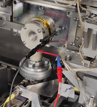
In addition, we have experience assisting previous users in developing and manufacturing custom electrochemistry in operando cells. Currently, there are a few models available at the beamline (see image below), but we are flexible enough to adapt to your sample and instrumental needs. Furthermore, I14 owns two different pieces of equipment for providing different potentials: Ivium OctoStat (https://www.ivium.com/product/octostat200/) and a Bio-potentiostat (https://www.stinstruments.com/electrochemistry/potentiostat-galvanostat-eis/sp-200-potentiostat/). Please  approach a member of the I14 team to discuss the operational details. Miguel Gomez Gonzalez will be pleased to hear from you to discuss electrochmistry experiments.

Custom made AMPIX electrochemistry cell
Example of custom-made AMPIX electrochemistry cell developed in collaboration with Prof. Grey group (Cambridge University).
Based on the original design published in Borkiewicz et al. (2012) Jornal of Applied Crystallography 45, 6, 1261–1269
(https://journals.iucr.org/paper?S0021889812042720).

Things to consider:

Sample preparation – You will need to apply your sample to a specific chip depending on which holder you choose. The size is limited to the space between the chips (approx. 2-8 microns).

Timescale – The timescale of change needs to correspond with our mapping times (e.g. too quick and you will miss it, too slow and you may run out of beamtime).

Testing – A certain amount of offline testing is usually beneficial to make an experiment successful.

Beam related effects – Some reactions can be induced by the X-ray beam

More info: https://doi.org/10.1080/09603409.2023.2213579.

  • Contact Us
  • About Diamond Light Source
  • Procurement
  • Supply Chain Transparency
  • Cookie Policy
  • Website Terms of Use
  • Privacy Notice

Diamond Light Source

Diamond Light Source is the UK's national synchrotron science facility, located at the Harwell Science and Innovation Campus in Oxfordshire.

Follow us on Facebook Follow us on Linkedin Follow us on Instagram Follow us on Bluesky

Diamond Light Source Ltd
Diamond House
Harwell Science & Innovation Campus
Didcot
Oxfordshire
OX11 0DE

See on Google Maps

Copyright © Diamond Light Source. Diamond Light Source® and the Diamond logo are registered trademarks of Diamond Light Source Ltd

Registered in England and Wales at Diamond House, Harwell Science and Innovation Campus, Didcot, Oxfordshire, OX11 0DE, United Kingdom. Company number: 4375679. VAT number: 287 461 957. Economic Operators Registration and Identification (EORI) number: GB287461957003.

feedback