• Diamond Home page
  • About
  • For Users
  • Public
  • Industry
  • Instruments
  • Careers
  • More Show more menu items
Search

About

  • About Diamond
  • About Synchrotrons
  • News and Features
  • Events
  • Contact Us
  • FAQ

For Users

  • Apply for beamtime
  • User guide
  • Diamond Users Commitee
  • FAQ
  • User Administration System
  • iSpyB

Industry

  • Techniques Available
  • Industry Research
  • Industry Case Studies
  • News
  • Meet the Industry Team

Science

  • Research
  • Computing
  • The Machine
  • Publications
  • Research Expertise
  • Membrane Protein Laboratory
  • Additional Facilities
  • Publications Database

Instruments

  • Biological Cryo-Imaging
  • Crystallography
  • Imaging and Microscopy
  • Macromolecular Crystallography
  • Magnetic Materials
  • Soft Condensed Matter
  • Spectroscopy
  • Structures and Surfaces

Diamond-II

  • Science
  • Machine
  • Beamlines
  • Software, Control and Computing
  • Infrastructure

Public

  • Public open days
  • School visits
  • Partner with Diamond
  • Explore Diamond

Procurement

  • Non-OJEU Tender Notices
  • OJEU PINs
  • OJEU Tender Notices
  • Registration Form

Careers

  • Vacancies
  • Info for applicants
  • Company Benefits
  • Apprenticeships
  • PhD Studentships
  • Work Placement

Main Content

Imaging and Microscopy
Sub-navigation
  • Imaging and Microscopy
  • I08
  • I08-1 (J08)
  • I12
  • DIAD
  • I13-1
  • ePSIC
  • I13-2
  • I14

In This Section

Sub Navigation
  • Welcome to I14
  • About I14
    • Beamline layout
    • Beamline instrumentation
    • Beamline techniques
      • Tips for the nanoprobe
      • X-ray Fluorescence (XRF)
      • X-ray Absorption Near Edge Structure (XANES)
      • Extended X-ray Absorption Fine Structure (EXAFS)
      • Sparse Scanning (XANES and EXAFS)
      • Differential Phase Contrast imaging (DPC)
      • X-ray diffraction (XRD)
      • Ptychography
      • Tomography
      • In situ/in operando
    • Laboratory equipment
  • I14 beamtime and access
  • Beamline research projects
  • Staff
  • Recent publications
  • Science Highlights and News
  • I14 Latest news
  • Accessibility Information

Instruments by Science Group

Macromolecular
Crystallography
Soft Condensed
Matter
Imaging and
Microscopy
Biological
Cryo-Imaging
Magnetic
Materials
Structures and
Surfaces
Crystallography
Spectroscopy

I14 Contact

I14 Control room:
Tel: +44 (0) 1235 778570

Principal Beamline Scientist: 
Majid Kazemian
Email: [email protected]
Tel: +44 (0) 1235 778222

Science Group Leader

Julia Parker

Email: [email protected]
Tel: +44 (0)1235 778924

I14 Hard X-ray Nanoprobe

Status: Operational

Beamsize: 50nm x 50nm
Energy: 5 - 20 keV

XAS: X-ray Absorption Spectroscopy XRF: X-ray Fluorescence Imaging X-ray Diffraction nXRD: nano X-ray Diffraction Ptychography Spectroscopy PCI: Phase Contrast Imaging Tomography Microscopy nSpectroscopy: nano Spectroscopy XANES: X-ray Absorption Near Edge Structure
  1. Instruments
  2. Imaging and Microscopy
  3. I14
  4. About I14
  5. Beamline techniques
  6. Tomography

Tomography

What it tells you: the above techniques, but in 3D!

Tomography allows us to collect XRF, DPC, ptychography or XRD information in three dimensions. This can help mitigate thickness or shape variations in your sample.

Tomography map

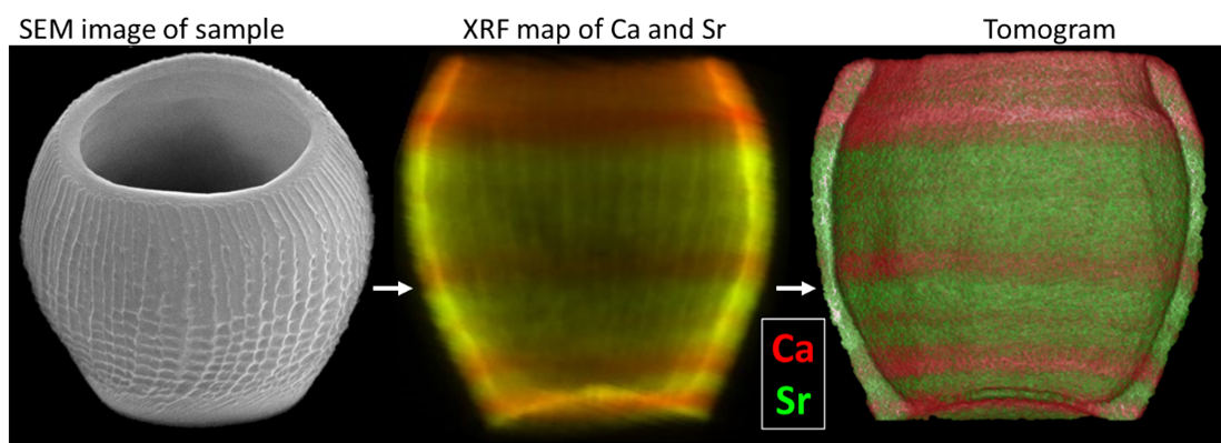
For this technique, we collect an image using the technique of your choice at angles from 0-180 degrees, usually at a rotational step size of 1-2 degrees. This leads to a lot of maps!

Tomography map sample

Things to consider:

Sample preparation – We require samples to be on pins, with nothing behind them in any direction. The pins need to be of a specific height and your sample size should be within a range of 20-30 microns. The larger your sample, the longer your map will be, and the less resolution you may have time for.  We are happy to discuss options for sample preparation.

Time – Tomography takes a long time (90 or 180 x the time for one map).

Data analysis – Our pipelines for analysis require some knowledge and input to work properly, and can be quite sample specific. You may need to iterate some steps to achieve the best alignment and reconstruction.  

More info: https://doi.org/10.1039/D3EM00509G.

  • Contact Us
  • About Diamond Light Source
  • Procurement
  • Supply Chain Transparency
  • Cookie Policy
  • Website Terms of Use
  • Privacy Notice

Diamond Light Source

Diamond Light Source is the UK's national synchrotron science facility, located at the Harwell Science and Innovation Campus in Oxfordshire.

Follow us on Facebook Follow us on Linkedin Follow us on Instagram Follow us on Bluesky

Diamond Light Source Ltd
Diamond House
Harwell Science & Innovation Campus
Didcot
Oxfordshire
OX11 0DE

See on Google Maps

Copyright © Diamond Light Source. Diamond Light Source® and the Diamond logo are registered trademarks of Diamond Light Source Ltd

Registered in England and Wales at Diamond House, Harwell Science and Innovation Campus, Didcot, Oxfordshire, OX11 0DE, United Kingdom. Company number: 4375679. VAT number: 287 461 957. Economic Operators Registration and Identification (EORI) number: GB287461957003.

feedback