• Diamond Home page
  • About
  • For Users
  • Public
  • Industry
  • Instruments
  • Careers
  • More Show more menu items
Search

About

  • About Diamond
  • About Synchrotrons
  • News and Features
  • Events
  • Contact Us
  • FAQ

For Users

  • Apply for beamtime
  • User guide
  • Diamond Users Commitee
  • FAQ
  • User Administration System
  • iSpyB

Industry

  • Techniques Available
  • Industry Research
  • Industry Case Studies
  • News
  • Meet the Industry Team

Science

  • Research
  • Computing
  • The Machine
  • Publications
  • Research Expertise
  • Membrane Protein Laboratory
  • Additional Facilities
  • Publications Database

Instruments

  • Biological Cryo-Imaging
  • Crystallography
  • Imaging and Microscopy
  • Macromolecular Crystallography
  • Magnetic Materials
  • Soft Condensed Matter
  • Spectroscopy
  • Structures and Surfaces

Diamond-II

  • Science
  • Machine
  • Beamlines
  • Software, Control and Computing
  • Infrastructure

Public

  • Public open days
  • School visits
  • Partner with Diamond
  • Explore Diamond

Procurement

  • Non-OJEU Tender Notices
  • OJEU PINs
  • OJEU Tender Notices
  • Registration Form

Careers

  • Vacancies
  • Info for applicants
  • Company Benefits
  • Apprenticeships
  • PhD Studentships
  • Work Placement

Main Content

Imaging and Microscopy
Sub-navigation
  • Imaging and Microscopy
  • I08
  • I08-1 (J08)
  • I12
  • DIAD
  • I13-1
  • ePSIC
  • I13-2
  • I14

In This Section

Sub Navigation
  • Welcome to I14
  • About I14
    • Beamline layout
    • Beamline instrumentation
    • Beamline techniques
      • Tips for the nanoprobe
      • X-ray Fluorescence (XRF)
      • X-ray Absorption Near Edge Structure (XANES)
      • Extended X-ray Absorption Fine Structure (EXAFS)
      • Sparse Scanning (XANES and EXAFS)
      • Differential Phase Contrast imaging (DPC)
      • X-ray diffraction (XRD)
      • Ptychography
      • Tomography
      • In situ/in operando
    • Laboratory equipment
  • I14 beamtime and access
  • Beamline research projects
  • Staff
  • Recent publications
  • Science Highlights and News
  • I14 Latest news
  • Accessibility Information

Instruments by Science Group

Macromolecular
Crystallography
Soft Condensed
Matter
Imaging and
Microscopy
Biological
Cryo-Imaging
Magnetic
Materials
Structures and
Surfaces
Crystallography
Spectroscopy

I14 Contact

I14 Control room:
Tel: +44 (0) 1235 778570

Principal Beamline Scientist: 
Majid Kazemian
Email: [email protected]
Tel: +44 (0) 1235 778222

Science Group Leader

Julia Parker

Email: [email protected]
Tel: +44 (0)1235 778924

I14 Hard X-ray Nanoprobe

Status: Operational

Beamsize: 50nm x 50nm
Energy: 5 - 20 keV

XAS: X-ray Absorption Spectroscopy XRF: X-ray Fluorescence Imaging X-ray Diffraction nXRD: nano X-ray Diffraction Ptychography Spectroscopy PCI: Phase Contrast Imaging Tomography Microscopy nSpectroscopy: nano Spectroscopy XANES: X-ray Absorption Near Edge Structure
  1. Instruments
  2. Imaging and Microscopy
  3. I14
  4. About I14
  5. Beamline techniques
  6. Ptychography

Ptychography

What it tells you: as with DPC you get an image of your overall sample, with added resolution

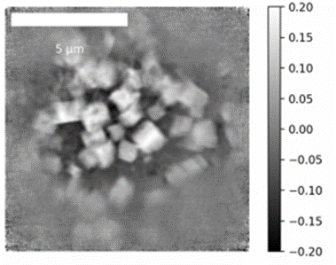
Ptychography uses overlapping measurements and computation to produce a higher resolution image. For this technique, we automatically place the sample at ~800 µm out of focus, generating a bigger (but less coherent) beam of ~1.5 µm 2 size.

This image is based on phase contrast and can be helpful for imaging structures that can be seen in DPC at higher resolution. Due to the mathematical retrieval algorithms used, the pixel size of the phase reconstructed image is not limited to the size of the beam, what may lead to the acquisition high resolution images, down to ~20 nm per pixel (energy dependent!).

Please note that ptychography is not a routine technique yet, but we encourage users with different samples to acquire some ptychography scans on their samples, should the DPC images described above look fine.

Things to consider:

Post-processing – Getting the best image can be highly sample specific. You may need to do some re-processing to optimise the computation. Nonetheless, we currently offer a “user friendly” system that automatically triggers different post-processing retrieval algorithms all at once. That allows the users to visualise several phase images in about ~1 hour after submission.

Signal – Too thick or too thin samples can result in a poor reconstruction.

Phase ptychography reconstruction
Example of a phase ptychography reconstruction unveiling cubical crystals of siderite (FeCO3) being formed as a consequence of iron corrosion.
  • Contact Us
  • About Diamond Light Source
  • Procurement
  • Supply Chain Transparency
  • Cookie Policy
  • Website Terms of Use
  • Privacy Notice

Diamond Light Source

Diamond Light Source is the UK's national synchrotron science facility, located at the Harwell Science and Innovation Campus in Oxfordshire.

Follow us on Facebook Follow us on Linkedin Follow us on Instagram Follow us on Bluesky

Diamond Light Source Ltd
Diamond House
Harwell Science & Innovation Campus
Didcot
Oxfordshire
OX11 0DE

See on Google Maps

Copyright © Diamond Light Source. Diamond Light Source® and the Diamond logo are registered trademarks of Diamond Light Source Ltd

Registered in England and Wales at Diamond House, Harwell Science and Innovation Campus, Didcot, Oxfordshire, OX11 0DE, United Kingdom. Company number: 4375679. VAT number: 287 461 957. Economic Operators Registration and Identification (EORI) number: GB287461957003.

feedback