Instruments by Science Group
I14 Contact
I14 Control room:
Tel: +44 (0) 1235 778570
Principal Beamline Scientist:
Majid Kazemian
Email: [email protected]
Tel: +44 (0) 1235 778222
Science Group Leader
Julia Parker
Email: [email protected]
Tel: +44 (0)1235 778924
I14 Hard X-ray Nanoprobe
Status: Operational
Beamsize: 50nm x 50nm
Energy: 5 - 20 keV
Energy: 5 - 20 keV
In-situ imaging
The nano X-ray techniques available at I14 can also be applied in a time resolved manner to image reactions, crystallization or other sample changes taking place in real time.
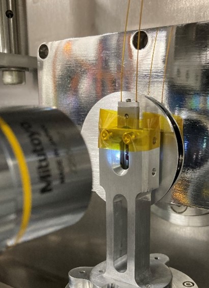
We have adapted in-situ TEM holders that can be placed on the beamline for imaging in either liquid or gas environments.
Samples can be heated or electrically biased, and the gas or liquid can be under constant flow or can flow in, and then be left to react.
- Two silicon nitride chips each with a window area are sandwiched together
- The heater chip is patterned to allow for either heating or biasing, and the top chip is unpatterned, but has a space for an o-ring for liquid sealing
- Liquid cell chips have small windows to reduce bulging, gas chip windows give a larger field of view
- They are clamped into the bespoke beamline holder so the windows on each chip align
- Capillary tubing and two holes in the heater chip allow flow through of either gas or liquid
Looped XRF, XANES or XRD scans can then be set up to follow a reaction within the cell.
Both DENS and Protochips in situ holders are available at the beamline.
Using the in-situ set up, we have looked at corrosion, catalysts and crystallization.
More information about catalysts in situ: A cell design for correlative hard X-ray nanoprobe and electron microscopy studies of catalysts under in situ conditions, J. Parker et al., J. Synchrotron Rad. 2022, 29, 431-438, https://doi.org/10.1107/S1600577521013576
Imaging in an in situ liquid environment: An in situ liquid environment for synchrotron hard X-ray nanoprobe microscopy, G. T. van de Kerkhof et al., 2023, Materials at High Temperatures, 40, 4, 371-375, https://doi.org/10.1080/09603409.2023.2213579

Diamond Light Source is the UK's national synchrotron science facility, located at the Harwell Science and Innovation Campus in Oxfordshire.
Diamond Light Source Ltd
Diamond House
Harwell Science & Innovation Campus
Didcot
Oxfordshire
OX11 0DE
Copyright © Diamond Light Source. Diamond Light Source® and the Diamond logo are registered trademarks of Diamond Light Source Ltd
Registered in England and Wales at Diamond House, Harwell Science and Innovation Campus, Didcot, Oxfordshire, OX11 0DE, United Kingdom. Company number: 4375679. VAT number: 287 461 957. Economic Operators Registration and Identification (EORI) number: GB287461957003.