Diamond Home Page
  • Diamond Home Page
  • About Us
  • For Users
  • Industry
  • Public
  • Science
  • Instruments
  • Careers
  • More
Search

On this website

About Diamond

  • About Diamond
  • About Synchrotron
  • News and Features
  • Events
  • Contact Us
  • FAQ

For Users

  • Apply for beamtime
  • User guide
  • Diamond Users Commitee
  • FAQ

Industry

  • Access Modes
  • Industry Research
  • Industry Case Studies
  • News
  • Meet the Industry Team

Science

  • Research
  • Computing
  • The Machine
  • Publications
  • Research Expertise
  • Membrane Protein Laboratory
  • Additional Facilities

Instruments

  • All the Instruments
  • Macromolecular Crystallography
  • Soft Condensed Matter
  • Imaging and Microscopy
  • Biological Cryo-Imaging
  • Magnetic Materials
  • Structures and Surfaces
  • Crystallography
  • Spectroscopy

Press Office

  • Media Enquiries
  • Media Resources
  • Scientists for Media Interview
  • Press Releases
  • Filming at Diamond

Careers

  • Vacancies
  • Info for applicants
  • Company Benefits
  • Apprenticeships
  • PhD Studentships
  • Work Placement

Public

  • News
  • How Diamond Works
  • Multimedia
  • Features
  • Visits Us
  • For School

Procurement

  • Non-OJEU Tender Notices
  • OJEU PINs
  • OJEU Tender Notices
  • Registration Form

Software & Tools

  • Publications Database
  • iSpyB
  • Diamond Wiki
  • User Administration System
  • Check the Machine Status

Social Diamond

  • Diamond Light Source on Twitter Follow us on Twitter
  • Diamond Light Source on Vimeo Watch us on Vimeo


  • Diamond Light Source on Facebook Follow us on Facebook
  • Diamond Light Source on LinkedIn Follow us on LinkedIn

Main Content

A brighter light for science
Sub-navigation
  • About Us
  • Governance
  • Legal & Compliance
  • Literature
  • News & Features
  • Events
  • Contact Us
  • Location

In This Section

Sub Navigation
  • Inside Diamond
  • Diamond News
  • Annual Review
    • Diamond Annual Review 2019
    • Diamond Annual Review 2018
      • Foreword
      • CEO Welcome
      • Beamline Development and Technical Summary
      • Science Group Updates
        • Macromolecular Crystallography Group
        • Surfaces and Interfaces Group
        • Magnetic Materials Group
        • Imaging and Microscopy Group
          • Update
          • Revealing the reactivity of iron in the Lena River basin
          • Investigating lithium ion battery failures
          • Corrective glasses provide sharper X-ray vision
          • Imaging multiphase fluid flow in permeable media
          • State-of-the-art electron microscopy offers clarity on diamond defects
        • Crystallography Group
        • Biological Cryo-Imaging Group
        • Spectroscopy Group
        • Soft Condensed Matter Group
      • Phase III Overview
      • Machine Operation and Development
      • Optics and Metrology Group
      • Detector Group
      • Data Analysis and Scientific Software Group
      • Data Acquisition Group
      • Key Facts and Figures
      • Collaborations
      • Industrial Liaison at Diamond
      • Engaging with Diamond Light Source
      • Governance and Management
      • Committee Membership
    • Diamond Annual Review 2017
    • Diamond Annual Review 2016
    • Diamond Annual Review 2015
    • Submission Guidelines
  • Literature Reviews
  1. Diamond Light Source
  2. Literature
  3. Annual Review
  4. Diamond Annual Review 2018
  5. Science Group Updates
  6. Imaging and Microscopy Group

Imaging and Microscopy Group

  • Sweet Success: Crystal structure of the full-length GLP-1 receptor bound to a peptide agonist

    Sweet Success: Crystal structure of the full-length GLP-1 receptor bound to a peptide agonist Jun 1, 2018

    To design such small molecules, a detailed map of the ...

  • Structural insights into the mechanism of bacterial ABC transporters

    Structural insights into the mechanism of bacterial ABC transporters Jun 1, 2018

    Bacteria utilise ATP-binding cassette (ABC) transporters ...

  • Structure of the surface layer surrounding Caulobacter crescentus bacteria

    Structure of the surface layer surrounding Caulobacter crescentus bacteria Jun 1, 2018

    Many types of single-celled microorganisms such as ...

  • How does an inspector call: structures of UGGT, the eukaryotic glycoprotein quality control checkpoint.

    How does an inspector call: structures of UGGT, the eukaryotic glycoprotein quality control checkpoint. Jun 1, 2018

    In all cells derived from animals, plants, fungi and ...

  • Insights into the mechanism of colistin resistance from crystal structures of MCR-1

    Insights into the mechanism of colistin resistance from crystal structures of MCR-1 Jun 1, 2018

    Gram-negative bacteria that are extensively resistant to ...

  • How human endoglin captures its ligand BMP9

    How human endoglin captures its ligand BMP9 Jun 1, 2018

    Endoglin (ENG)/CD105 is a mammalian glycoprotein that is ...

Paul Quinn, Science Group Leader

Update

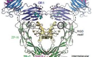
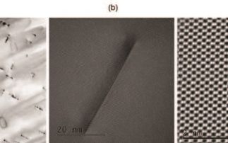
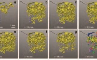
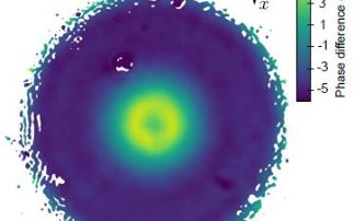
Imaging and Microscopy is a newly formed science group, which brings together eight experimental facilities (I08, J08, DIAD, I12, I13-1, I13-2, I14 and ePSIC) with a new imaging group leader to coordinate and drive the group’s activities. These facilities use electrons and X-rays to image samples under different experimental conditions across a diverse range of length scales and time scales. Different contrast mechanisms allow for imaging of sample properties such as elemental composition, density and structure and this ability to extract image sample properties in minute detail lends itself to a wide range of scientific areas from chemistry and catalysis to environmental science, materials science, biology, medicine, and cultural heritage. Read more ...

Highlights

  • Revealing the reactivity of iron in the Lena River basin

    Revealing the reactivity of iron in the Lena River basin Jun 1, 2018

    With the melting Arctic ice cap causing its rivers to ...

  • State-of-the-art electron microscopy offers clarity on diamond defects

    State-of-the-art electron microscopy offers clarity on diamond defects Jun 1, 2018

    Flawless diamonds are extremely rare and expensive. Most ...

  • Imaging multiphase fluid flow in permeable media

    Imaging multiphase fluid flow in permeable media Jun 1, 2018

    Knowing how fluids move through micron-sized voids (or ...

  • Corrective glasses provide sharper X-ray vision

    Corrective glasses provide sharper X-ray vision Jun 1, 2018

    The intense X-ray light produced by modern synchrotron ...

  • Investigating lithium ion battery failures

    Investigating lithium ion battery failures Jun 1, 2018

    The 18650 cylinder is the most widely used lithium ion ...

  • Contact Us
  • About Diamond Light Source
  • Careers
  • Procurement
  • Legal notices & Cookie policy
  • Supply Chain Transparency

Diamond Light Source

Diamond Light Source is the UK's national synchrotron science facility, located at the Harwell Science and Innovation Campus in Oxfordshire.

Copyright © 2020 Diamond Light Source


Diamond Light Source Ltd
Diamond House
Harwell Science & Innovation Campus
Didcot
Oxfordshire
OX11 0DE

See on Google Maps

Diamond Light Source® and the Diamond logo are registered trademarks of Diamond Light Source Ltd