Corrective glasses provide sharper X-ray vision
Jun 1, 2018
Jun 1, 2018
The intense X-ray light produced by modern synchrotron radiation sources and X-ray free-electron lasers (XFELs) allows the fine structure and dynamics of matter to be studied in exquisite detail. In principle, beams should be able to be focused to a few nanometres and below. Such small intense X-ray nanobeams are crucial to concentrate the radiation onto a given sample. However, the beam’s short wavelength places stringent requirements on today’s X-ray optics, which limit resolution due to refractive, diffractive, or reflective distortions (known as aberrations) of the resulting image.
The ever-increasing brightness of modern storage ring sources and XFELs enables studying the structure and dynamics of matter with unprecedented spatial and temporal resolution. To fully benefit from the power of these sources, it is crucial to confine the X-ray beam and concentrate the radiation onto the sample. This is essential to improve not only the spatial resolution and sensitivity of X-ray analytical techniques, but also to generate highly intense X-rays that facilitate the generation of extreme states of matter and enable the study of non-linear X-ray phenomena. However, this requires X-ray optics with high numerical aperture (NA) that can withstand the extremely bright pulses of modern sources. Unfortunately, suitable X-ray optics are limited by fabrication technology, and trade-offs need to be made between aberration-free performance and highest possible NA.
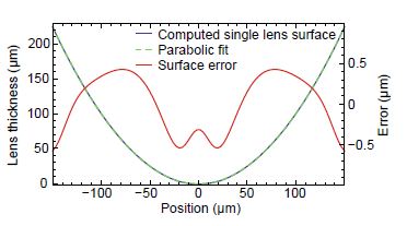
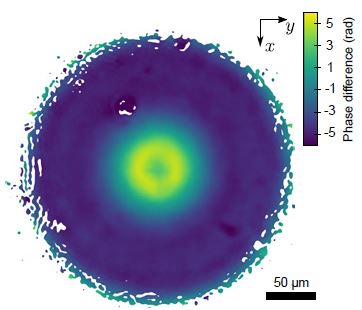
Ptychography1, an X-ray scanning coherent diffraction microscopy technique, has evolved to one of the most important methods for X-ray beam and optics characterisation. Here, a nano-structured sample, positioned in the vicinity of the focal plane was scanned across the beam while far-field diffraction patterns were recorded with appropriate spatial overlap. With the help of elaborate phase retrieval algorithms2 the complex wavefield at the sample position could be retrieved unambiguously. From these data the wavefront error in the exit pupil of the focusing optics was calculated with highest sensitivity and spatial resolution3 (Fig. 1). For focusing a stack of 20 beryllium compound refractive lenses (Be CRLs) with a radius of curvature of 50 μm and a geometrical aperture of 300 μm was used at an X-ray energy of 8.2 keV. The rotational symmetric phase error shown in Fig. 1, a clear signature for spherical aberration, originates from tiny but repetitive shape errors of individual Be CRLs within the large lens stack. To reproduce the wavefield error in Fig. 1, numerical simulations have been performed where the lens shape deviation from an ideal parabola was iteratively refined. The retrieved deformation of an individual lens surface is shown in Fig. 2. As Be CRLs are fabricated via a coining process by pressing two parabolic stamps into a Be foil, an improvement of the already very small deformation error of 500 nm by a significant amount appears very challenging.

Instead, a general scheme to correct for residual aberrations in any X-ray optics was employed. By introducing corrective glasses into the optical path -- tailor-made to the specific optics -- true diffraction-limited focusing could be achieved. From the phase error in Fig. 1 a corrective phase plate was modelled that corrects the wavefield deformation by introducing an opposing phase shift. Fig. 3 shows the phase plate made out of fused silica by ultrashort-pulse laser ablation4.
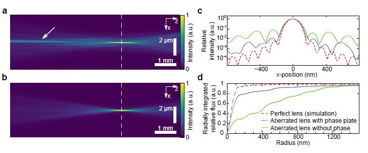
Due to spherical aberration, the nanofocus using the uncorrected optics was spoiled by X-rays being partly focused further upstream, highlighted by the arrow in Fig. 4a. This led to unwanted radiation around the focal spot, shown by the solid green line in the focal plane profile of Fig. 4c. Compared to a perfect lens (dashed red line in Fig. 4c), intensities in side lobes were increased by over an order of magnitude. In practice, these side lobes reduce the attainable resolution in X-ray microscopy applications due to an effective spot size broadening, illustrated by the radially integrated intensity profile in Fig. 4d. In addition, the peak intensity in the main focal spot is significantly reduced, as most of the radiation is distributed in the outer lobes.
By installing the tailor-made glasses behind the Be CRLs, the performance of the optics was strongly improved. With the suppression of spherical aberration, most X-rays were now focused within the same focal plane, illustrated by the clean beam caustic in Fig. 4b. This resulted in lowered intensities by an order of magnitude in lobes surrounding the focal spot, shown by the dotted blue line in Fig. 4c, and ultimately a reduced effective focal spot size as shown in Fig. 4d. The corrective glasses transformed the Be CRL optics from an aberrated system into a diffraction-limited hard X-ray optics. While the uncorrected optics could only focus less than 30 % of the radiation compared to the ideal optics (Strehl ratio below 0.3), the corrected focus gathered more than 85 % (Strehl ratio 0.85). At the same time the geometrical aperture and NA of the optics is unaffected.
With this novel scheme, many X-ray optics can be improved in performance beyond current manufacturing limitations. The principle is not only applicable to refractive optics as shown here, but can also be transferred to reflective and diffractive optics as well. As the glasses are very compact and easy to align, they can also be retrofitted to existing beamline optics. This benefits not only the resolution and sensitivity in classical X-ray microscopy schemes, but may also lead to new opportunities for isochoric X-ray heating and non-linear X-ray optics at XFEL sources.
Funding acknowledgement: This work was supported by the German Ministry of Education and Research (BMBF) under Grant Number 05K13OD2, the DFG under Grant SCHR 1137/1-1 and the Swedish Research Council. Frank Seiboth, Andreas Schropp and Christian Rödel acknowledge funding from a Peter Paul Ewald fellowship of the Volkswagen Foundation. Parts of this research were carried out at the coherence branch of beamline I13 at Diamond Light Source and beamline P06 at PETRA III at DESY, a member of the Helmholtz Association. Use of the Linac Coherent Light Source, SLAC National Accelerator Laboratory, is supported by the US Department of Energy, Office of Science, Office of Basic Energy Sciences under Contract No. DE-AC02-76SF00515. The MEC instrument is supported by the US Department of Energy, Office of Science, Office of Fusion Energy Sciences under Contract No. SF00515.
Seiboth F, Schropp A, Scholz M, Wittwer F, Rödel C, Wünsche, M, Ullsperger T, Nolte S, Rahomäki J, Parfeniukas K, Giakoumidis S, Vogt U, Wagner U, Rau C, Boesenberg U, Garrevoet J, Falkenberg G, Galtier EC, Lee HJ, Nagler B, Schroer CG. Perfect X-ray focusing via fitting corrective glasses to aberrated optics. Nature Communications 8, 14623, doi:10.1038/ncomms14623 (2017).

Diamond Light Source is the UK's national synchrotron science facility, located at the Harwell Science and Innovation Campus in Oxfordshire.
Diamond Light Source Ltd
Diamond House
Harwell Science & Innovation Campus
Didcot
Oxfordshire
OX11 0DE
Copyright © Diamond Light Source. Diamond Light Source® and the Diamond logo are registered trademarks of Diamond Light Source Ltd
Registered in England and Wales at Diamond House, Harwell Science and Innovation Campus, Didcot, Oxfordshire, OX11 0DE, United Kingdom. Company number: 4375679. VAT number: 287 461 957. Economic Operators Registration and Identification (EORI) number: GB287461957003.