Paul Quinn, Science Group Leader
Imaging and Microscopy is a newly formed science group, which brings together eight experimental facilities (I08, J08, DIAD, I12, I13- 1, I13-2, I14 and ePSIC) with a new imaging group leader to coordinate and drive the group’s activities. These facilities use electrons and X-rays to image samples under different experimental conditions across a diverse range of length scales and time scales. Different contrast mechanisms allow for imaging of sample properties such as elemental composition, density and structure and this ability to extract image sample properties in minute detail lends itself to a wide range of scientific areas from chemistry and catalysis to environmental science, materials science, biology, medicine, and cultural heritage.
The Scanning X-ray Microscopy (SXM) beamline (I08) is for morphological, elemental and chemical speciation on a broad range of organic-inorganic interactions in a 250 - 4400 eV photon energy range, and sample investigations under ambient or cryogenic conditions. I08 has a range of applications including biological and biomedical sciences, earth and environmental science, geochemistry, and materials science. During the reporting period, the performance for lower photon energies on I08 was improved significantly, in particular for carbon Near Edge X-ray Absorption Fine Structure (NEXAFS) spectromicroscopy. I08 faced its first major upgrade as a result of the Scanning and Mapping projects. This upgrade is now finalised and offers users superior performance in data collection and software interfaces to the user community. The design and construction of a soft X-ray spectro- and tomo-ptychography branchline (J08) progresses according to plan. This instrument is expected to be available for experiments in the second half of 2019.
The Dual Imaging and Diffraction (DIAD) beamline (K11) will be the first beamline to offer two X-ray microscopy techniques (imaging and diffraction) applied synchronously with a switching time of a few milliseconds. This enables in situ structural characterisation experiments taking advantage of both techniques simultaneously.
DIAD is being built to use light from a ten pole permanent magnet wiggler. The diffraction technique is conducted using monochromatic light, whereas the imaging technique can be performed with monochromatic or polychromatic (‘pink’) beam. The X-ray energy can be chosen separately for both techniques in the range from 7 - 38 keV. The beamline is under construction and is expected to start commissioning activities in the middle of 2019. First users are expected by the end of that year. The user-community is already heavily engaged in many aspects of beamline operation, particularly in the selection and specification of dedicated sample environments. A mechanical test-rig for diffraction and tomography will be one of the main instruments to allow in situ experiments for a variety of scientific disciplines like engineering and material science, bio-materials and hard tissues, geology and mineralogy, and soilplant interactions. The beamline will also aim to provide sample environments for other communities such as energy, electro-chemistry and corrosion science.
The I12 (JEEP) beamline uses a 4.2 T superconducting wiggler to provide polychromatic and monochromatic X-rays in the energy range 50 - 150 keV. The high photon energies provide good penetration through large or dense samples. The beamline offers beam sizes ranging from 50 x 50 microns for diffraction, up to 90 x 25 mm for imaging. These beam characteristics enable the study of materials and processes inside sample environments without unacceptable attenuation of the beam, using macro-scale samples that are more representative of the process under study. X-ray techniques available are radiography, tomography, energy-dispersive diffraction, monochromatic and white-beam 2D diffraction/scattering and small-angle X-ray scattering. The beamline’s two flexible experimental hutches allow users to bring their own rigs and sample chambers. I12 has a diverse user community (materials science and engineering; chemical processing; biomedical engineering; geoscience; environmental science; physics, palaeontology) who make full use of the beamline’s versatility. On the technical side, I12 has commissioned a new IR lamp furnace (“Helios”), designed for diffraction and imaging experiments on small samples and capillaries at temperatures > 1000o C. In the optics hutch, there is a new heat load management filter which removes photons below 50 keV. The new rotating disc design replaces the original fixed SiC filter design, which was unreliable. The rotating filter was developed and tested over an extended period to ensure robustness and reliability. This was achieved without disrupting beamline user operations.
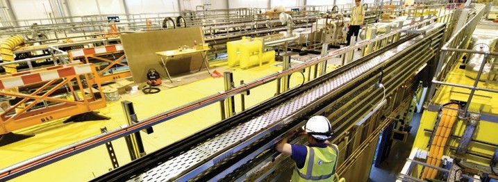
- Figure 1: Construction of the hutches nearing completion for DIAD.
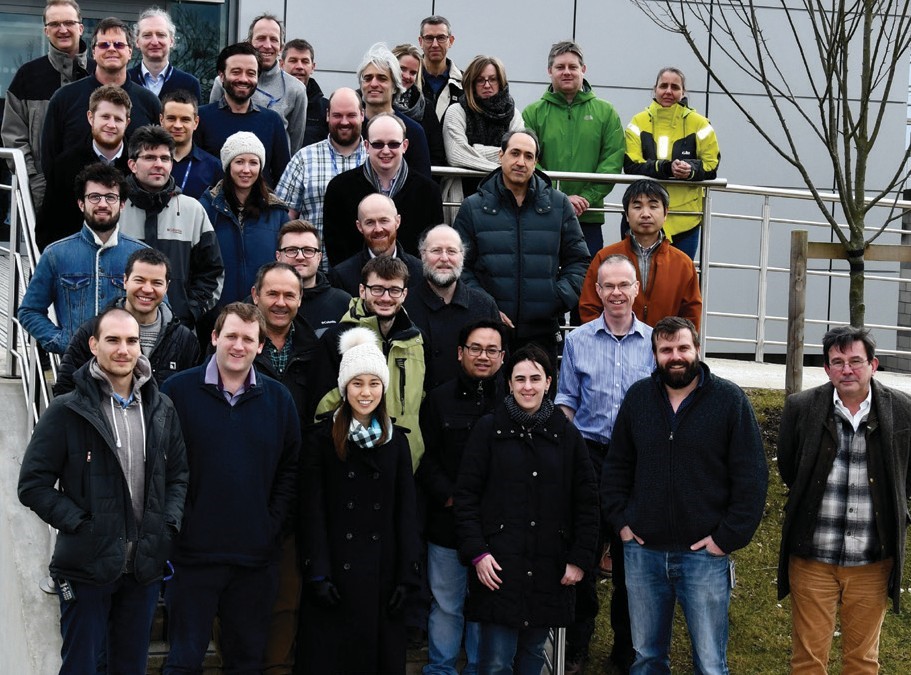
- Figure 2: The scientists, engineers, technicians and PDRAs that make up the Imaging and Microscopy Group.
The I13 imaging and coherence beamline aims for multiscale imaging in the energy range of 6 - 30 keV. The achievable resolution ranges from several microns to some tens of nanometers with two branchlines operating independently for this purpose. The Diamond-Manchester imaging branchline performs mainly in-line phase contrast tomography with a strong emphasis on dedicated sample environments. Failure of lithium batteries, material cracks, the structure of ice cream, bones under load, the storage of CO2 in brine and sandstone are some examples of the studies conducted under realistic conditions. Two projects are currently under development for submicron and phase sensitive imaging. A new full-field microscope will perform Zernike phase contrast imaging over a field of view of 50-100 μm and a resolution of 50 - 100 nm and a grating interferometry setup will provide superb image quality, measuring the absolute phase and providing small angle information allowing us to identify nano-sized structures from micrometer resolution X-rays. The highest spatial resolution, of 30 nm,is achieved on the coherence branch with ptychographic imaging. Our most important development is that we are now able to use the EXCALIBUR photon counting detector at 50 Hz frame rate which has reduced ptycho-tomography scans from days to a few hours. We can now routinely perform ptychography as a standard user experiment, enabling the beamline’s ambition for multi-scale imaging. A large number of pilot experiments are currently underway, many of which previously used the imaging branch but can now exploit higher resolution ptychographic imaging. Examples are the study of battery failure, the origin and structure of particles from the Fukushima accident, or the micro- and nano-structure of insects.
I14, the Hard X-ray Nanoprobe beamline, welcomed its first users in March 2017 which kicked off an exciting year of commissioning, developments and experiments. I14 offers a small beam of 100 - 200 nm for high resolution imaging. Over the last year I14 has offered X-ray fluorescence, diffraction and XANES mapping with spatial resolutions down to 100 nm. Thermal spray coatings, corroded metal surfaces, meteorites, metallic particles in cells, photovoltaic films and radionuclide particles are just a sample of the many science areas and successful experiments conducted so far. The beamline is still in its optimisation phase and over the coming year the spatial resolution will improve to ~50 nm and new techniques and facilities such as ptychography, cryogenic sample handling and in situ sample environments will be rolled out for routine use.
In 2017 the Electron Physical Sciences Imaging Centre (ePSIC) at Diamond welcomed its first users. The two transmission electron microscopes which make up the centre, a JEOL ARM 200 and a JEOL GRAND ARM 300, were brought to Diamond through collaboration with Johnson Matthey and Oxford University. The ARM 200 is a state-of-the-art probe-corrected analytical microscope capable of atomic resolution electron energy loss and X-ray spectroscopy. The ARM 300 is a dedicated imaging instrument aligned across a wide range of accelerating voltages (30 - 300 keV). It is both probe- and imaging-corrected and has numerous detectors including a fast direct electron detector (operating at up to 2000 fps). These combined capabilities make this a unique resource for electron microscopy within the UK. With in situ sample holders, users at ePSIC can perform variable temperature measurements from 100 to 1600 Kelvin to directly image the atomic structure of materials during thermally driven transitions. This in situ capability will be expanded upon over the coming year. An Oxford Instruments EDX detector has been added to the ARM 300 to allow combined X-ray spectroscopy and high-resolution imaging. The state of the art instrumentation available at ePSIC has attracted both established electron microscopists looking to develop new techniques and scientists with limited previous electron microscopy experience interested in the atomic structure of their samples. The collaboration of the expert staff at ePSIC with this range of users is helping to bring cutting edge microscopy techniques to the wider material science community.

Diamond Light Source is the UK's national synchrotron science facility, located at the Harwell Science and Innovation Campus in Oxfordshire.
Diamond Light Source Ltd
Diamond House
Harwell Science & Innovation Campus
Didcot
Oxfordshire
OX11 0DE
Copyright © Diamond Light Source. Diamond Light Source® and the Diamond logo are registered trademarks of Diamond Light Source Ltd
Registered in England and Wales at Diamond House, Harwell Science and Innovation Campus, Didcot, Oxfordshire, OX11 0DE, United Kingdom. Company number: 4375679. VAT number: 287 461 957. Economic Operators Registration and Identification (EORI) number: GB287461957003.