Instruments by Science Group
I22 Contact
Beamline phone numbers:
+44 (0) 1235 77 8617
+44 (0) 1235 77 8713
Principal Beamline Scientist:
Nick Terrill
Tel: +44 (0) 1235 778047
E-mail: [email protected]
Science Group Leader
Robert Rambo
Email: [email protected]
Tel: +44 (0)1235 56 7675
The main role for this end station set up is to carry out mapping and dynamic experiments on heterogeneous samples with regions of interest in few tens of micron size range. Most microfocus experiments carried out on I22 so far have used beamsize between 10-25 micron. Previously these experiments have been carried out at 14keV to ease attenuation due to air scatter. Phase 1 of the BCO Upgrade offers significantly enhanced capability with variable beamsize, energy and evacuated flight tubes to improve data quality.
The new set-up provides demagnification and focusing of the primary beam down to a minimum spot size of approximately 10 μm2. The microfocus sample stages are still available for accurate sample positioning and with the completion of the BCO upgrade in late 2019 a new inline microscipe capability for ALL modes will be available.
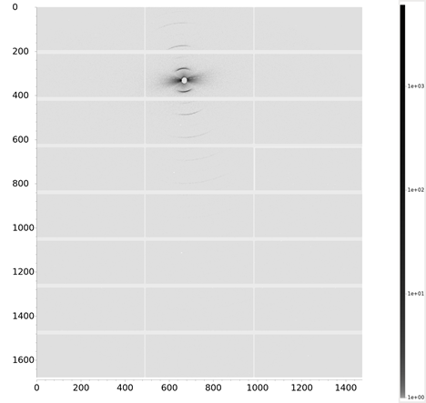
The new optical layout has significantly reduced divergence over the old end station which gives access to much lower q than was available previously. While we currently offer energies in the range 14-20 keV, the new set up using the SAXS camera tubes allows us to consider on a case by case basis lower energy microfocus experiments.
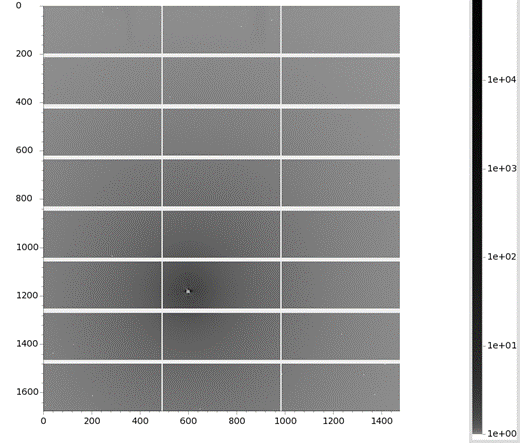
We have recorded a flux of 5.6 x 10¹⁰ photons/sec in the focused beam at 14 keV, very similar to the old system but with significantly reduced backgrounds, hugely improved q-range and lower divergence.
 |  |
This option is now available to users throughout the schedule, via the normal PRP route.
Latest Microfocus Publication
Understanding how anisotropic fibre-symmetric materials work is important for biological research and bioinspired design. We introduce an analytical model for X-ray diffraction in nanofibrillar materials with fibre symmetry that considers arbitrary diffraction angles, 3D orientation, strain heterogeneity, and angular misalignment. Validated by scanning synchrotron WAXD on mantis shrimp cuticle, our model accurately reconstructs fibre textures and detects gradients in orientation, strain, and dispersion. By fitting multiple reflections, we improve parameter extraction, especially in complex regions. This framework advances WAXD analysis in heterogeneous fibre materials and can be integrated with tomographic or machine-learning methods for efficient structural characterization when sample rotation is limited.
Anisotropic diffraction of materials with fibre symmetry: application to chitin cuticle
Wang, Y., Snow, T., Terrill, N. & Gupta, H.S.
IUCrJ, 2026, Issue 1
DOI : 10.1107/S2052252525009686

Diamond Light Source is the UK's national synchrotron science facility, located at the Harwell Science and Innovation Campus in Oxfordshire.
Diamond Light Source Ltd
Diamond House
Harwell Science & Innovation Campus
Didcot
Oxfordshire
OX11 0DE
Copyright © Diamond Light Source. Diamond Light Source® and the Diamond logo are registered trademarks of Diamond Light Source Ltd
Registered in England and Wales at Diamond House, Harwell Science and Innovation Campus, Didcot, Oxfordshire, OX11 0DE, United Kingdom. Company number: 4375679. VAT number: 287 461 957. Economic Operators Registration and Identification (EORI) number: GB287461957003.