Instruments by Science Group
Contact
eBIC admin enquiries
Tel: +44 (0) 1235 56 7480
Email: [email protected]
Office hours: 08.30-16.30(UK) Mon-Fri
Science enquiries
Peijun Zhang
Tel: +44 (0) 1235 77 8878
Email: [email protected]
Daniel Clare
Tel: +44 (0) 1235 56 7501
Email: [email protected]
Yuriy Chaban
Tel: +44 (0) 1235 77 8207
Email: [email protected]
Christos Savva
Tel: +44 (0) 1235 77 8976
Email: [email protected]
Vojtech Pražák
Tel: +44 (0) 1235 77 8131
Email: [email protected]
Training enquiries
Lorna Malone
Tel: +44 (0) 1235 394182
Email: [email protected]
Science Group Leader (interim)
Daniel Clare
Email: [email protected]
Tel: +44 (0) 1235 56 7501
Funded by a grant from


eBIC
Status: Operational
Accelerating Voltages: 3-30, 200 & 300 kV
Cryo fluorescence microscope
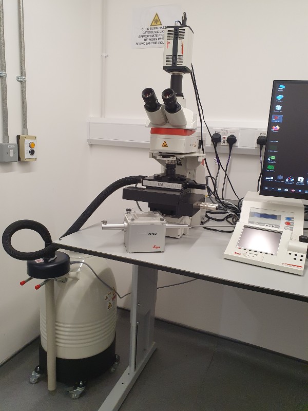
The cryoCLEM is a dedicated cryo-fluorescence microscope, designed to maintain cryo-conditions for specimen handling.
It provides high resolution imaging to enable the user to target the regions of interest for the correlative process.
The Correlative Light and Electron Microscopy (CLEM) process includes the extra information provided by the fluorescence signal and guides the cryo-EM tilt series collection, making the data acquisition much more accurate and efficient.
cryoCLEM is designed and optimised for the cryo-correlation workflow.
It features a specially designed cryo-stage that goes with the optical microscope, an easy-to-use grid transfer system, as well as a special software that is easy to use.
Features include
Cryo-stage
A built in cryo-stage provides exceptional mechanical stability, a sealed sample compartment minimises ice contamination, and temperature monitoring with automatic LN2 pumping ensures the safety of the sample.
Grid transferring components
The grid transfer shuttle minimises ice contamination, keeps the temperature stable and makes grid handling an easy process.
Fully automated acquisition
The grid mapping is fully automated at chosen location(s). It is capable of full-grid mapping that includes automatic lateral tiling, multi axial heights with auto-focusing, multi-colour channels, etc.
Image processing
Image projection, stitching and overlaying is performed automatically. The result can be exported into an EM software compatible format. The image post processing functions such as 3D deconvolution can further improve the resolution via computational processing.
Optical component
Specially designed high numerical aperture objective (NA = 0.9) with long working distance is optimised for cryogenic temperature imaging. The add on of a sCMOS camera insures the sensitivity to capture weak signal, as well large field of view.
Imaging modes
-
transmission, to visualise ice thickness, cracks and crystalline ice
-
reflection mode to show the surface of the grid
-
fluorescence mode
cryo-CLEM filter cubes
| Filter system | Excitation | Dichroic | Emission |
| DAPI ET | 350/50 | 400 | 460/50 |
| GFP ET | 470/40 | 495 | 525/50 |
| TXR ET | 560/40 | 585 | 630/75 |
| Far red Y5 | 620/60 | 660 | 700/75 |
cryoCLEM instrument manager

Diamond Light Source is the UK's national synchrotron science facility, located at the Harwell Science and Innovation Campus in Oxfordshire.
Diamond Light Source Ltd
Diamond House
Harwell Science & Innovation Campus
Didcot
Oxfordshire
OX11 0DE
Copyright © Diamond Light Source. Diamond Light Source® and the Diamond logo are registered trademarks of Diamond Light Source Ltd
Registered in England and Wales at Diamond House, Harwell Science and Innovation Campus, Didcot, Oxfordshire, OX11 0DE, United Kingdom. Company number: 4375679. VAT number: 287 461 957. Economic Operators Registration and Identification (EORI) number: GB287461957003.