Instruments by Science Group
I12 Contact
Science Group Leader
Julia Parker
Email: [email protected]
Tel: +44 (0)1235 778924
I12 JEEP: Joint Engineering, Environment and Processing
Status: Operational
Beamsize: From 50 micrometer x 50 micrometer to 50 mm x 15 mm (EH1) or 100 mm x 30 mm (EH2)
Energy: 53 – 150 keV
Energy: 53 – 150 keV
The beamline I12 - JEEP is capable of delivering white or monochromatic beam of a large size and a high flux to be used for diffraction and imaging experiments.
| Source | Super-conducting wiggler, 4.2 T, 48 mm periodicity, 22 full field periods |
| Beam modes | White beam or monochromatic beam |
| Monochromator | Si (111) cryo-cooled double crystal bent Laue |
| Maximum beam size and divergence | EH1: 50 mm (H) × 13 mm (V) EH2: 94 mm (H) × 26 mm (V) corresponding to 1 mrad × 0.3 mrad angular beam size permitted throughout the beamline |
| Energy range: | 53 - 150 keV |
| Photon flux (EH1, 53 keV at 300 mA ring current) | 1.8 × 10¹¹ photons s-1 mm-2 per 0.1% bandwidth |
| Relative energy resolution: | From 10-3 to 2 × 10⁻⁴, depending on monochromator setting |
BEAMLINE LAYOUT
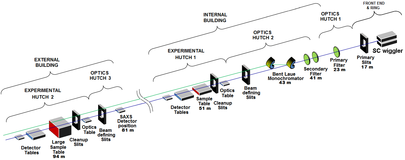
Optical and functional schematic of the I12 - JEEP beamline.
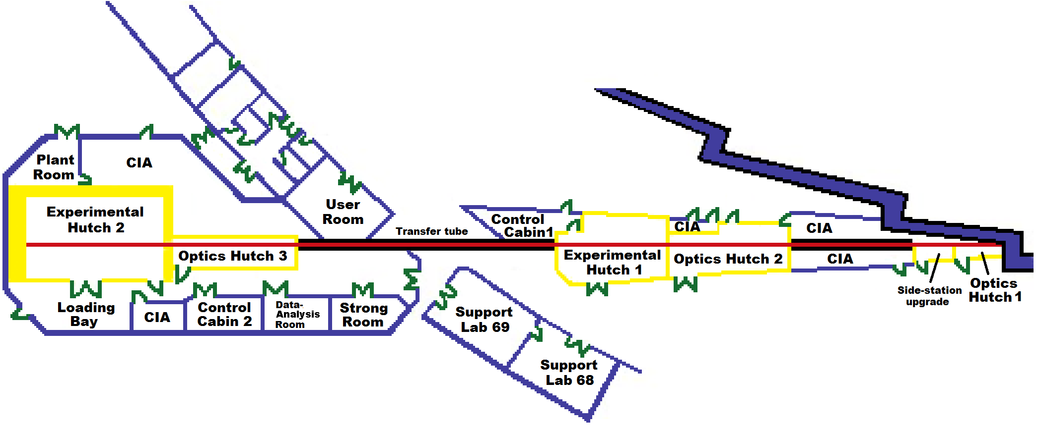
Building schematic of the I12 - JEEP beamline
The I12 beamline is divided into two parts, with an Internal Building and an External Building
Internal Building includes:
· Optics Hutch 1 (OH1)
· Side station upgrade
· Optics Hutch 2 (OH2)
· Experimental Hutch 1 (EH1)
· Control Cabin 1 (CC1) for control of the internal beamline
· User room (UR) to provide additional space for users and beamline staff using CC1
External Building includes:
· Optics Hutch 3 (OH3)
· Experimental Hutch 2 (EH2)
· Control Cabin 2 (CC2) for control of the external beamline
· Data-Analysis Room (DAR)
· Strong Room for storage of valuable samples
Peripheral labs at the perimeter of the experimental hall:
· Support Lab 68
· Support Lab 69
BEAMLINE SUMMARY
Source
· Super-conducting multipole wiggler
· Constant peak field: 4.2 T
· Number of pole pairs at full field: 22
· Period length: 48 mm
· K value: 18.8
· Critical energy: 25 keV
· 1 mrad × 0.3 mrad divergence permitted into beamline
Optics
· Bent crystal Laue monochromator using silicon (111) crystal; variable bending radius for both crystals independently from flat to 90 m with concave side oriented towards the source; asymmetry angle 44o; monochromatic beam at fixed vertical offset of 50 mm above white beam; liquid N2 cooling on first crystal; second crystal cooled by Cu-braid connected to liquid N2 line
· Permanent filters reduce the heat load on optics and attenuate x-rays with energies below 50 keV: 2 × 1.1 mm CVD diamond filter, 1× 4 mm CVD SiC filter. The first diamond filter acts as window to separate beamline vacuum from the storage ring
· Optional Cu filters with selectable thickness from 1 to 12 mm
· Beam defining slits for EH1 at 47 m
· Beam defining slits for EH2 at 88 m
Calculated white beam and monochromatic beam intensities at the sample position can be downloaded here.
Experimental Hutch 1 (EH1)
EH1 is located inside the experimental hall. It contains:
· Optics table (for instruments to be placed before the sample)
· Sample table (for samples and sample environments; maximum weight is 50 kg for tomography and 200 kg for radiograpy and diffraction)
· Detectors. Detectors are mounted on automated tables: on the Large Detector Table or on the Small Detector Table. Detectors may be remotely positioned and exchanged during an experiment, allowing different techniques to be applied to the same sample
For Diffraction
- Large 2D area diffraction detector. The sample-to-detector distance can be varied from 500 mm to 2400 mm or set to 4500 mm; large transverse movement of detector through Experimental Hutch is possible
For Imaging:
- X-ray camera with selectable optics to remotely change the field of view and the resolution. Four modules offer fields of view between 3 mm and 50 mm width. Two interchangeable back-end detectors are available for either high resolution or high speed measurements: PCO.edge for high resolution imaging and Vision Research MIRO 310M for high speed imaging
· Additional services: single phase and three phase power, cooling water, compressed air, pressurized gases, manual crane, exhaust system
Experimental techniques in EH1
All techniques are performed at high x-ray energies according to the energy range of the beamline
· Static and time-resolved radiography and tomography
· Static and time-resolved powder diffraction using monochromatic X-rays
· Single crystal diffraction using monochromatic X-rays
· Diffuse scattering on crystalline or amorphous materials (single crystal diffuse scattering, pair-distribution function analysis “PDF”)
· Small angle scattering at the sample-to-detector distance of 4.5 m covering q-range from 0.03 1/Å to 0.8 1/Å for energy of 55 keV.
Some experimental techniques can be combined during the same visit, on the same sample. For details please read here.
Experimental Hutch 2 (EH2)
EH2 is located in an external building. It contains:
· Optics table (for instruments to be placed before the sample)
· Sample table (for samples and sample environments with weight up to 2000 kg)
· Detectors. Detectors are mounted on automated tables: on Large Detector Table or on Small Detector Table. Detectors may be remotely positioned and exchanged during an experiment, allowing different techniques to be applied to the same sample
For Diffraction:
- Large 2D area diffraction detector. The sample-to-detector distance can be varied from 1000 mm to 2600 mm or set to 6300 mm; large transverse movement of detector through Experimental Hutch is possible
- Energy dispersive X-ray detector Canberra with 23 high purity Ge crystals in a semi-circular arrangement
For Imaging:
- X-ray camera with selectable optics to remotely change the field of view and the resolution. Four modules offer fields of view between 3 mm and 50 mm width. Two interchangeable back-end detectors are available for either high resolution or high speed measurements: PCO.edge for high resolution imaging and Vision Research MIRO 310M for high speed imaging
- Large Field of View camera for tomography of large samples; incorporates two high-resolution imaging detectors PCO.edge providing field of view approx. 104 × 46 mm
· INSTRON servo-hydraulic 100 kN uniaxial mechanical test rig for in situ and ex situ experiments. Rig is also available for “offline” work such as sample preparation before beamtime
· Additional services (single phase and three phase power, cooling water, compressed air, pressurized gases, manual crane, exhaust system, loading bay for vehicle access)
Experimental techniques in EH2
All techniques are performed at high x-ray energies according to the energy range of the beamline
· Static and time-resolved radiography and tomography
· Static and time-resolved powder diffraction using monochromatic X-rays
· Time-resolved energy dispersive powder diffraction
· Single crystal diffraction using monochromatic X-rays with restricted goniometry
· Diffuse scattering on crystalline or amorphous materials (single crystal diffuse scattering, pair-distribution function analysis “PDF”)
· Small angle scattering at the sample-to-detector distance of 6300 mm covering q-range from 0.02 1/Å to 0.5 1/Å for energy of 55 keV.
Some experimental techniques can be combined during the same visit, on the same sample. For details please read here.
Sample environments
· Cryostream (100 - 473 K).
· Custom designed ODISC heating furnace (25 - 800 oC) - for powder diffraction experiments and special imaging experiments; furnace does not include the flow cell, so the possible oxidation of sample during the heating in air must be taken into account.
· Linkam heating furnace (25 - 1300 oC) - for powder diffraction experiments or radiography experiments (not suitable for tomography experiments); Linkam furnace allows high-temeprature measurements in Argon flow or in vacuum (up to 10-3mbar).
· Custom designed metal heating block (25 - 95 oC) - for powder diffraction experiments during chemical synthesis.
· Custom designed Helios heating furnace (200 - 1300 oC) - for powder diffraction and special imaging experiments in air in transmission geometry.
· INSTRON 100 kN uniaxial servo-hydraulic mechanical test rig (available only in Experimental Hutch 2).
· Cooling water supply.
· Compressed air (max. pressure 5 bar).
· Pressurized He, Ar, CO2, N2 of technical quality (max. pressure 10 bar).
Support laboratory
· Zeiss Axiotech Vario reflected light microscope (with possibility to save images)
· Zeiss Stemi-2000 stereo light microscope (with possibility to save images)
· Fume cupboard
· Small balances (mg-g)
· Small fridge (2-5 oC)
· Laboratory oven ED115 (30-300 oC), download manual
· Ultrasonic bath
· De-ionized water supply
· Compressed air and pressurized gases of technical quality
· Manual goniometers
· 3-phase power
· 1-phase high current (32 A) power
· Strong room for secure sample and sample environment storage

Diamond Light Source is the UK's national synchrotron science facility, located at the Harwell Science and Innovation Campus in Oxfordshire.
Diamond Light Source Ltd
Diamond House
Harwell Science & Innovation Campus
Didcot
Oxfordshire
OX11 0DE
Copyright © Diamond Light Source. Diamond Light Source® and the Diamond logo are registered trademarks of Diamond Light Source Ltd
Registered in England and Wales at Diamond House, Harwell Science and Innovation Campus, Didcot, Oxfordshire, OX11 0DE, United Kingdom. Company number: 4375679. VAT number: 287 461 957. Economic Operators Registration and Identification (EORI) number: GB287461957003.