Instruments by Science Group
I14 Contact
I14 Control room:
Tel: +44 (0) 1235 778570
Principal Beamline Scientist:
Majid Kazemian
Email: [email protected]
Tel: +44 (0) 1235 778222
Science Group Leader
Julia Parker
Email: [email protected]
Tel: +44 (0)1235 778924
I14 Hard X-ray Nanoprobe
Status: Operational
Beamsize: 50nm x 50nm
Energy: 5 - 20 keV
Energy: 5 - 20 keV
Project 1: NANOMATERIALS TRANSFORMATION IN ENVIRONMENTAL SOLUTIONS
With the continued escalating of industrial application and production of engineered nanomaterials (ENMs), there arises the responsibility for understanding, mitigating, and reducing the ecological risks of these nanoscale entities. Specifically, their incorporation into commercial goods, followed by their disposal, may have unexpected environmental consequences. While there are concerns regarding the environmental ramifications of the increase of ENMs production, real evidence remains deficient for robust risk assessments. Predominantly, research projects have studied pristine (raw) materials, with only a small number of works studying the fate, persistence, translocation across mediums, and the temporal kinetics of ENMs. It has been stablished that the physicochemical characteristics of ENMs can undergo considerable modifications, contingent upon the encountered environments (e.g., via processes such as agglomeration, aggregation, speciation, and varied chemical transformations). In ecological contexts, ENMs undergo dissolution, modulate their surface chemistries (e.g., through ligand binding), aggregate, undergo chemical transformations, and release ions.
Consequently, it is pivotal to accurately characterise the materials before use, in different environments and at the nanomaterial-environment interface, to evaluate their environmental impact. In addition, there is an urgent need for studies which combine meticulous particle characterisation and sophisticated imaging techniques under realistic experimental scenarios. Paired with bioactivity assays into organisms, this information could provide a greater insight into ENMs behaviour and a better appreciation of potential effects on the environment.
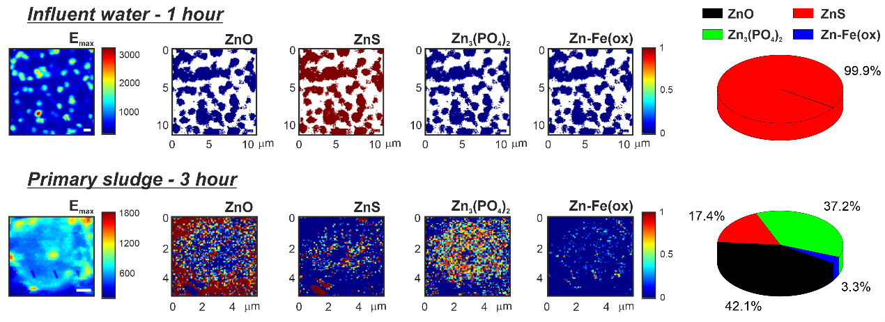
With a beam size of about ~50 nm, the hard X-ray nanoprobe (I14 beamline) at Diamond offers the capability to monitor dynamic changes to the morphology and surface chemistry of the ENMs in situ within relevant hydrated environments, and with an outstanding combination of energy and spatial resolution, by X-ray fluorescence (XRF) microscopy. In addition, the chemical speciation of the transformed species can be interrogated by X-ray spectroscopy near-edge structure (XANES) analysis afterwards (Fig. 1).
Figure 1 - XRF microscopy data of ZnO nanorods
- Figure 1 - XRF microscopy of ZnO nanorods
Figure 1.- XRF microscopy data of ZnO nanorods incubated in influent water for 1 hour (top) and in primary sludge for 3 hours (bottom). [Left] Fluorescence image acquired at the maximum of Zn K-edge (Emax = 9669 eV). [Middle] Speciation maps were calculated by XANES for the expected Zn- species: ZnO, ZnS, Zn3(PO4)2, and Zn adsorbed to Fe-oxyhydroxides (Zn-Fe(ox)) (from left to right), where the red colour equals a 100% contribution, and the blue colour corresponds to 0% (being the white pixels pure background – no-Zn detected). [Right] Pie charts with the average of all individual (pixel-by-pixel) percentages were also generated, representing the main contribution of each Zn-species. All the scale bars (white and black) are equal to 1 mm. © 2021 The Authors. Advanced Sustainable Systems published by Wiley-VCH GmbH. This is an open access article under the terms of the Creative Commons Attribution License, which permits use, distribution and reproduction in any medium, provided the original work is properly cited: Gomez-Gonzalez et al. Adv. Sustainable Syst. 2021, 2100023 (DOI: 10.1002/adsu.202100023).
Project 2: THE HIDDEN PATHWAYS OF (SMALL) PLASTIC POLLUTION
At Diamond Light Source, we are committed to deliver world class science and enhance our research activities that benefits society and the economy. A recent project conducted by researchers at the UK’s national synchrotron facility on its Hard X-ray Nanoprobe (I14 beamline), looked at how microplastics waste may interact with zinc oxide (ZnO) nanomaterials in both freshwater and seawater scenarios. Their work published in Global Challenges and echoed in Labmate, revealed worrying environmental implications for aquatic organisms in the food chain.
In recent decades, there has been a dramatic increase in the manufacture of engineered nanomaterials (tiny, tiny particles about 1000 times thinner than a human hair), which has inevitably led to their environmental release. Similarly, Zinc oxide (ZnO) is among the more abundant nanomaterials fabricated due to its advantageous use in electronics, semiconducting, and for antibacterial purposes. At the same time, plastic waste has become ubiquitous and may break down into smaller pieces called microplastics. These also are tiny, but ~100 times bigger than the nanomaterials. Because both these components are getting disposed more often, Dr Miguel Gomez-Gonzalez et al. decided to study their fate when they are potentially being combined in freshwater and oceans and to help make environmental risk assessments more accurate.
The ability of zinc oxide, both pure nanomaterials and those released from a sunscreen, to stick to very small pieces of plastic has big implications. These plastics can even come from everyday items like exfoliating facial cleansers. In this study, we found the microplastics can carry even smaller particles of zinc from place to place. As a consequence, fish or other aquatic organisms could swallow these microplastics, ingesting zinc particles at the same time.
In a broader effort to investigate the fate of even smaller pieces of plastics, we have teamed up with Dr Nathaniel Clark - University of Plymouth, to become project partners of ADVANCE: ADvanced Visualisation And Nano-scale Characterisation of Exposure.
Micro- and nanoplastics are increasingly found in human organs, including the liver. However, a critical barrier to understanding their health impact is our inability to reliably see these particles inside human tissues. Unlike metal-based nanoparticles, whose cellular trafficking and clearance are well understood, nanoplastics remain effectively invisible under most analytical systems.
This project will develop the first multimodal, label-free nanoscale imaging workflow to visualise plastic particles in human liver models. By integrating advanced imaging methods with spatial biology approaches, this research will map when, where and how micro- and nanoplastics interact with liver cells, allowing us to understand their biological significance and potential contribution to liver disease. The ultimate goal is seeing the Invisible, by understanding how plastics enter human tissues.
Preliminary research has localised europium-labelled nanoplastics (Eu-NPs) in macrophage cells by TEM microscopy, inside membrane-bound compartments (Fig. a). Complementary measurements at the I14 beamline further highlighted areas of Eu accumulation (Fig. b). However, these approaches cannot verify its chemical identity—an essential requirement, especially when analysing real-world samples such as biopsies, where other particles will be present.
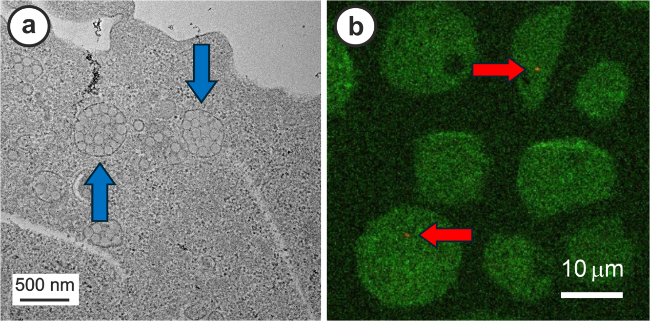
- Figure 2. Europium-labelled nanoplastics (Eu-NPs) in macrophage cells by TEM microscopy, inside membrane-bound compartments
Figure: a) Suspected Eu NP internalisation inside of macrophage cells following a 24-hour exposure, where nanoplastic clusters surrounded by membrane vesicles were revealed by TEM analysis (blue arrows). b) Nano XRF map measured at I14 showing the Eu-signal (orange colour dots, signalled by red arrows) over the K-signal for the macrophage (green colour). Note, the Eu-label is roughly 1-8% of the particle mass.

Diamond Light Source is the UK's national synchrotron science facility, located at the Harwell Science and Innovation Campus in Oxfordshire.
Diamond Light Source Ltd
Diamond House
Harwell Science & Innovation Campus
Didcot
Oxfordshire
OX11 0DE
Copyright © Diamond Light Source. Diamond Light Source® and the Diamond logo are registered trademarks of Diamond Light Source Ltd
Registered in England and Wales at Diamond House, Harwell Science and Innovation Campus, Didcot, Oxfordshire, OX11 0DE, United Kingdom. Company number: 4375679. VAT number: 287 461 957. Economic Operators Registration and Identification (EORI) number: GB287461957003.