Instruments by Science Group
Contact
eBIC admin enquiries
Tel: +44 (0) 1235 56 7480
Email: [email protected]
Office hours: 08.30-16.30(UK) Mon-Fri
Science enquiries
Peijun Zhang
Tel: +44 (0) 1235 77 8878
Email: [email protected]
Daniel Clare
Tel: +44 (0) 1235 56 7501
Email: [email protected]
Yuriy Chaban
Tel: +44 (0) 1235 77 8207
Email: [email protected]
Christos Savva
Tel: +44 (0) 1235 77 8976
Email: [email protected]
Vojtech Pražák
Tel: +44 (0) 1235 77 8131
Email: [email protected]
Training enquiries
Lorna Malone
Tel: +44 (0) 1235 394182
Email: [email protected]
Science Group Leader (interim)
Daniel Clare
Email: [email protected]
Tel: +44 (0) 1235 56 7501
Funded by a grant from


eBIC
Status: Operational
Accelerating Voltages: 3-30, 200 & 300 kV
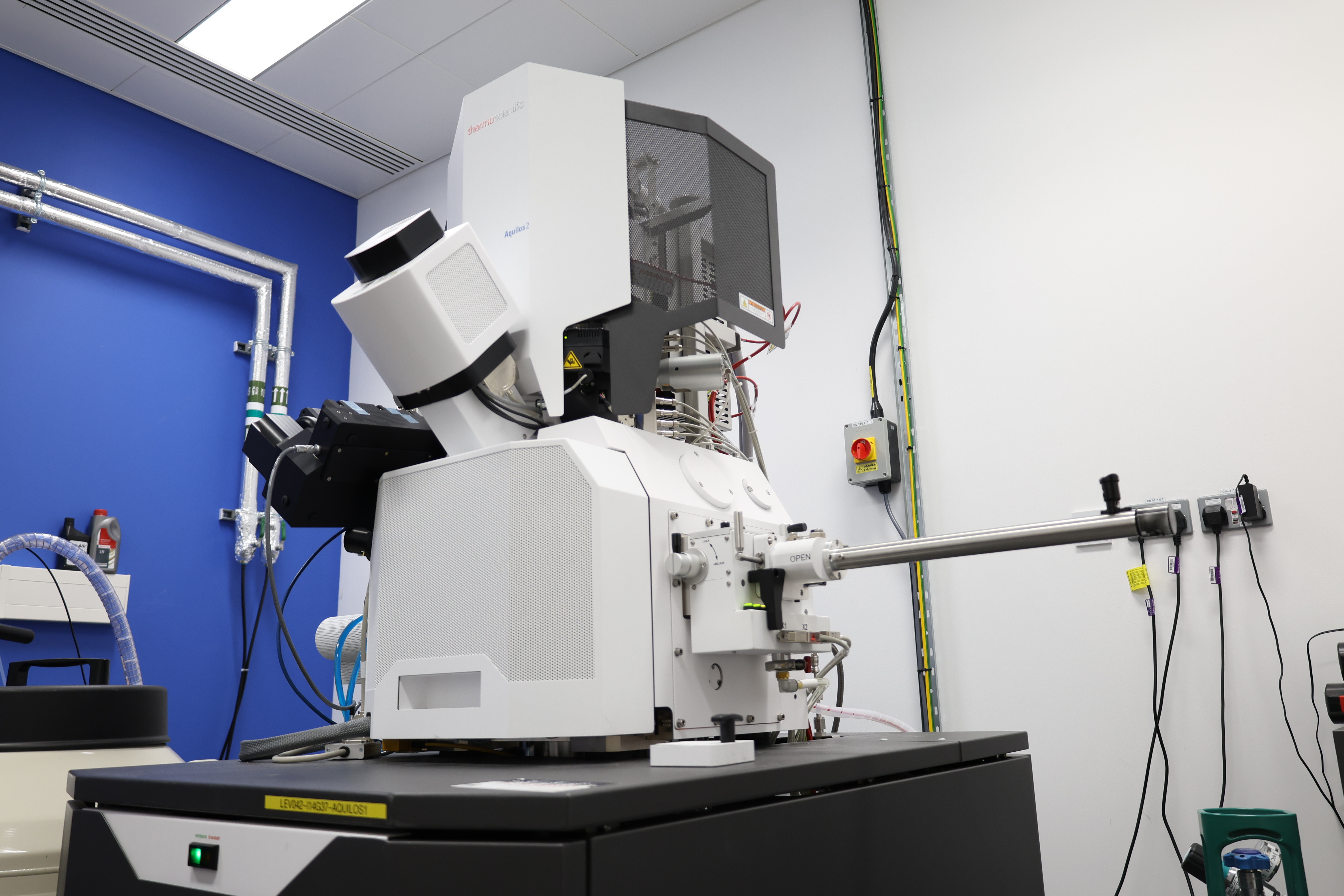
Scios and Aquilos cryoFIB/SEMs
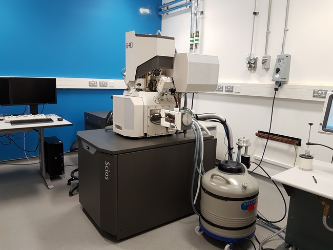
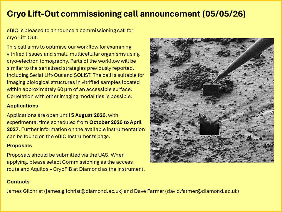
Focused ion beam/scanning electron microscopes (FIB/SEMs) allow for nanoscale milling of vitrified biological specimens, and mainly produce electron transparent lamellae but are also capable of directly imaging cellular features.
Lamellae are analysed using cryo-Electron Tomography (cryo-ET) which can reveal high-resolution biological structures in their close-to-native environment and direct imaging can provide cellular context.
These powerful techniques have allowed researchers to move toward a molecular-scale understanding of living systems.
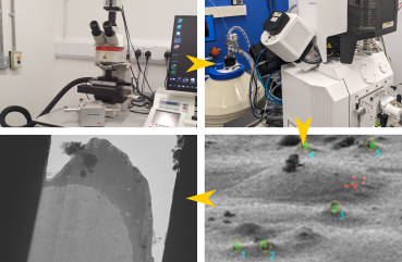
The Scios and Aquilos cryoFIB/SEMs form part of the multimodal imaging and sample preparation workflow available at Diamond, including cryo-fluorescent microscopy and high-resolution TEM imaging via one of eBIC’s four Titan Krios.
All of the systems in eBIC’s multimodal imaging pipeline are capable of accepting AutoGrid rings, for seamless transfer between different imaging modalities.
FIB/SEM technological advances have increased throughput and milling precision, yielding higher quality results in less time, making these systems ideal for examination of vitrified biological material at cryogenic temperatures.
Chamber-mounted enhancements
For both systems, sputter coating with metallic platinum is available and can improve imaging stability before milling. Sputter coating on lamellae can also improve phase plate imaging in the transmission electron microscope.
The gas injection system, integrated within the main chamber of both systems, coats frozen specimens with an organoplatinum compound to protect sensitive biological material from the ion beam and homogenise milling rates.
The Aquilos has two additional chamber-mounted options: a micromanipulator (EasyLift) and a fluorescent microscope (Meteor).
The EasyLift is an in-chamber, cryo-micromanipulator which allows lamellae from vitrified biological specimens e.g. tissues, to be extracted for thinning to electron transparency.
The Meteor is an in-chamber solution for identifying fluorescent biological targets in cells, tissue, vitreous ice and lamellae. The Meteor can act as a stand-alone correlative module, but also complements the cryoCLEM [link to: eBIC Instruments - cryoCLEM] at eBIC.
cryoFIB/SEMs specifications
|
|
Scios |
Aquilos |
|
e-beam energy (keV) |
0.2 - 30 |
0.2 - 30 |
|
Electron Source |
Schottky thermal FEG |
Schottky thermal FEG |
|
Ion-beam energy (keV) |
0.5 – 30 |
0.5 – 30 |
|
Ion source |
Ga liquid metal |
Ga liquid metal |
|
Cryo-transfer |
Quorum PP3010 |
ThermoFisher Scientific |
|
Stage rotation (°) |
No |
360 |
|
Sputter coating |
Incorporated into the PP3010 |
Within main chamber |
|
Grids per load |
Two |
Two |
|
Detectors |
ETD (SE) |
ETD (SE, BSE) T1 (in-lens, BSE) T2 (in-lens, BSE) |
FEG – Field emission gun, ETD – Everhart-Thornley detector, SE – Secondary electrons,
BSE – Back-scattered electrons, SI – Secondary ions
Meteor specifications
|
Objective |
Olympus Fluorite, 50x (WD 1.0mm, NA 0.80) |
|
Lightsource |
Omicron LedHub
|
|
Filters |
|
|
Camera |
Andor Sona - sCMOS, 6.5 μm pixel size |
CryoFIB and correlative milling
Submit your proposal
- lamella preparation on frozen cells and microcrystals
- automated, on-grid milling only
- Rapid and BAG access
- fluorescent microscopy correlation
For more information please see here.
Scios and Aquilos instrument manager

Diamond Light Source is the UK's national synchrotron science facility, located at the Harwell Science and Innovation Campus in Oxfordshire.
Diamond Light Source Ltd
Diamond House
Harwell Science & Innovation Campus
Didcot
Oxfordshire
OX11 0DE
Copyright © Diamond Light Source. Diamond Light Source® and the Diamond logo are registered trademarks of Diamond Light Source Ltd
Registered in England and Wales at Diamond House, Harwell Science and Innovation Campus, Didcot, Oxfordshire, OX11 0DE, United Kingdom. Company number: 4375679. VAT number: 287 461 957. Economic Operators Registration and Identification (EORI) number: GB287461957003.