Instruments by Science Group
I23 Contact
Beamline Phone Number:
+44 (0) 1235 567406
Principal Beamline Scientist:
Armin Wagner
Tel: +44 (0) 1235 778560
E-mail: [email protected]
Science Group Leader
Dave Hall
Email: [email protected]
Tel: +44 (0) 1235 778926
I23 Long-Wavelength MX
Status: Operational in optimisation mode
Wavelength: 1.1 – 5.9 Å
Energy: 2.1 – 11 keV
Energy: 2.1 – 11 keV
![]()
Overview
I23 complements the existing suite of seven MX beamlines at Diamond being optimised for operation in the wavelength range from 1.1 to 5.9 Å. The in-vacuum experimental end station minimizes absorption and scattering effects. A large semi-cylindrical detector allows measurements of a large range of diffraction angles and a multi-axis goniometer is available for crystal alignment and orientation. An X-ray tomography setup is integrated into the beamline end station to obtain the crystal shape and volume as a basis of an analytical absorption correction.
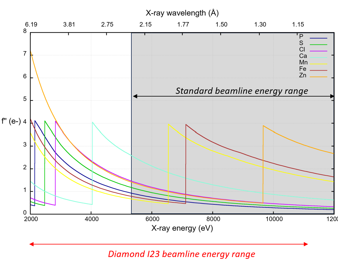
Variation of coefficient f’’ with X-ray wavelength (or energy) showing typical absorption K-edges accessible at the I23 beamline.
Light atoms identification and location by anomalous scattering
More than a third of all known proteins bind metal ions. Metal ions play key roles in a broad range of cellular processes, they are involved in protein structure stability and catalysis; with traditional examples of zinc fingers in transcription factors and iron in haemoglobin. Therefore, identifying metal ion-binding sites is important for understanding the biological functions of proteins and further helps in designing potent therapeutics.
The unique wavelength range of the macromolecular crystallography beamline I23 at Diamond Light Source allows identification and location of metal ions and lighter atoms of biological relevance (Ca, K, S, P and Cl) using X-ray anomalous scattering in crystal structure analysis.
In a typical experiment, anomalous datasets are collected at two wavelengths, above and below the ion or element absorption edge, and then processed to calculate phased anomalous difference Fourier maps. The difference in anomalous peak heights between these two datasets allows the direct identification and localisation of the ion in the protein structure. We successfully used this method in different projects to experimentally map ions in crystal structures.
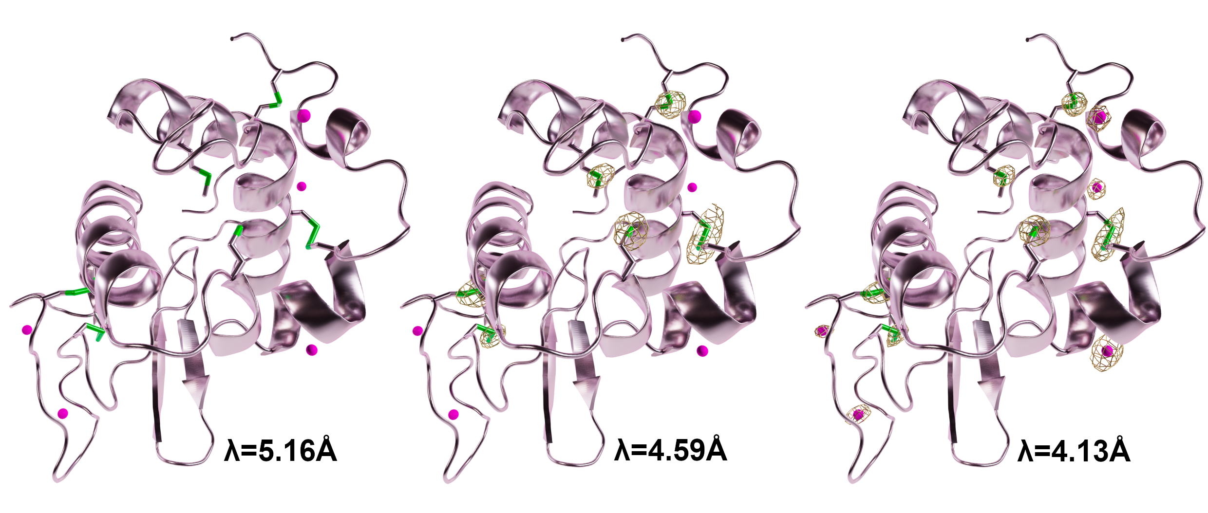
Identification of chlorine and sulfur atoms in Lysozyme structure
Experimental phasing by native-SAD
Initially, anomalous scattering was primarily used for experimental phasing, but with the recent advances of AlphaFold, this technique is used less frequently. However, in some cases, such as when predicted models are unavailable or unsuitable for molecular replacement, experimental phasing can provide unbiased electron density maps. Additionally, experimental phasing can be combined with molecular replacement to improve the quality of initial electron density maps and accelerate model building, which is especially useful in difficult projects.
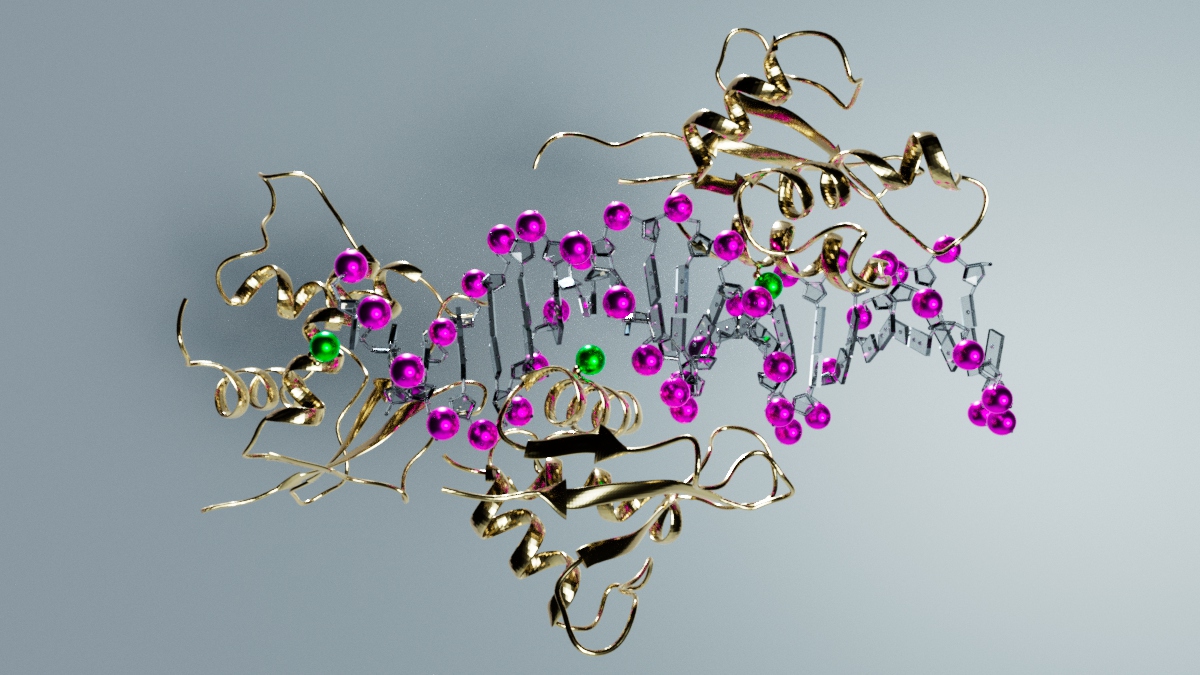
Nucleic acid:protein complexes structures can be solved using the anomalous signal from sulfur and phosphorus.
I23 beamline reference papers
K. El Omari, I. Forsyth, R. Duman, C.M. Orr et al. Utilizing anomalous signals for element identification in macromolecular crystallography. Acta Cryst. D (2024).
Y. Lu, R. Duman, J. Beilsten-Edmands, G. Winter et al. Ray-tracing analytical absorption correction for X-ray crystallography based on tomographic reconstructions. J Appl Crystallogr. (2024).
K. El Omari, R. Duman, V. Mykhaylyk, C.M. Orr et al. Experimental phasing opportunities for macromolecular crystallography at very long wavelengths. Commun Chem. (2023).
A. Wagner, R. Duman, K. Henderson, V. Mykhaylyk. In-vacuum long-wavelength macromolecular crystallography Acta Cryst. D72 (2016).

Diamond Light Source is the UK's national synchrotron science facility, located at the Harwell Science and Innovation Campus in Oxfordshire.
Diamond Light Source Ltd
Diamond House
Harwell Science & Innovation Campus
Didcot
Oxfordshire
OX11 0DE
Copyright © Diamond Light Source. Diamond Light Source® and the Diamond logo are registered trademarks of Diamond Light Source Ltd
Registered in England and Wales at Diamond House, Harwell Science and Innovation Campus, Didcot, Oxfordshire, OX11 0DE, United Kingdom. Company number: 4375679. VAT number: 287 461 957. Economic Operators Registration and Identification (EORI) number: GB287461957003.