• Diamond Home page
  • About
  • For Users
  • Public
  • Industry
  • Instruments
  • Careers
  • More Show more menu items
Search

About

  • About Diamond
  • About Synchrotrons
  • News and Features
  • Events
  • Contact Us
  • FAQ

For Users

  • Apply for beamtime
  • User guide
  • Diamond Users Commitee
  • FAQ
  • User Administration System
  • iSpyB

Industry

  • Techniques Available
  • Industry Research
  • Industry Case Studies
  • News
  • Meet the Industry Team

Science

  • Research
  • Computing
  • The Machine
  • Publications
  • Research Expertise
  • Membrane Protein Laboratory
  • Additional Facilities
  • Publications Database

Instruments

  • Biological Cryo-Imaging
  • Crystallography
  • Imaging and Microscopy
  • Macromolecular Crystallography
  • Magnetic Materials
  • Soft Condensed Matter
  • Spectroscopy
  • Structures and Surfaces

Diamond-II

  • Science
  • Machine
  • Beamlines
  • Software, Control and Computing
  • Infrastructure

Public

  • Public open days
  • School visits
  • Partner with Diamond
  • Explore Diamond

Procurement

  • Non-OJEU Tender Notices
  • OJEU PINs
  • OJEU Tender Notices
  • Registration Form

Careers

  • Vacancies
  • Info for applicants
  • Company Benefits
  • Apprenticeships
  • PhD Studentships
  • Work Placement

Main Content

Macromolecular Crystallography
Sub-navigation
  • MX
  • I03
  • I04
  • I04-1
  • I23
  • I24
  • VMXi
  • VMXm
  • HeXI
  • XFEL-HUB
  • XChem
  • MPL
  • Crystallisation Facility
  • MX Data Analysis
  • Staff Research
  • Contacts

In This Section

Sub Navigation
  • MX Responsive Beamtime Guide
  • Manual
    • Preparing for Beamtime
    • ISPyB/SynchWeb
    • Sample Holders
    • Shipping Samples
    • Remote Access
    • Data Collection Software
      • Navigating in the GUI
      • User Options
      • Baton Panel
      • Message of the Day
      • Load a Sample
      • Manually Mounted Samples
      • Sample Alignment
      • Centering Methods
      • Grid Scan
      • X-ray Centring
      • Fluorescence Data
      • Data Collection
      • Anomalous Data Collection
        • Inverse Beam SAD
        • Wedged MAD
      • Command Queue
      • Line Scan
      • Default File Names
      • Tools
      • Annealing Crystals
      • Washing Crystals
      • The HC1 Perspective
    • Data Analysis
    • Data Backup
    • Webcams
    • Acknowledging MX beamlines
    • Introduction to Eiger2 X Detectors for MX at Diamond
    • Redhat user environment
  • Science Highlights
  • Useful Calculators
  • MX beamtime guide for COVID-19 work
  • MX Training
  • Getting Started at Diamond MX

Instruments by Science Group

Macromolecular
Crystallography
Soft Condensed
Matter
Imaging and
Microscopy
Biological
Cryo-Imaging
Magnetic
Materials
Structures and
Surfaces
Crystallography
Spectroscopy
  1. Instruments
  2. MX
  3. Common
  4. Manual
  5. Data Collection Software
  6. Anomalous Data Collection
  7. Inverse Beam SAD

Inverse Beam SAD

What is an Inverse Beam SAD data collection?

The circle on the figure below represents 360o sweep on the same crystal with the same energy. For example, GDA will first collect sweep 1 (0 to 45o), then sweep 2  at 180o apart (181 to 225o), followed by sweep 3 (46 to 90o) and so on...

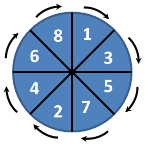
The advantage using inverse beam SAD data collection compared to regular SAD data is that the Friedel pairs will be collected with equivalent dose. It will result in a better assesment of the anomalous signal less polluted by radiation damage.

How to set an inverse beam SAD data collection

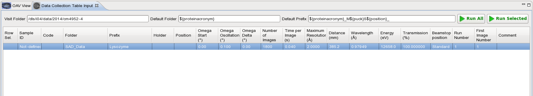
In GDA, go to "Data Collection" then click on the tab "Data Collection Table Input"

Note: Click on the images to view full size

Enter the parameters required for your data collection. For example omega start 0 degree, 0.1o oscillation, 0.040s exposure, energy at 12658 eV and 1800 images for total oscillation of 180o. You need to be aware that after converting into inverse beam SAD mode the total oscillation will doubled and be effectively 360o in this case. 


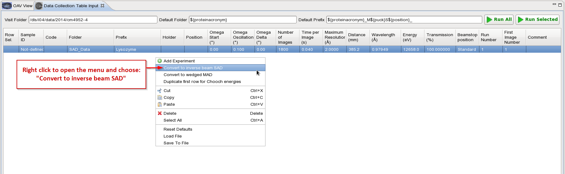
Then convert entry into inverse beam SAD as shown below:

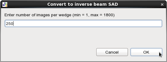
In this example we choose to do wedge of 25o (250 images with 0.1o oscillation). Note that for inverse beam SAD data collection we recommend to use 5o  wedge or lower.

The data collection has been broken up into 25o wedges and reorganized so that each wedge is collected 180o apart. The prefix name has also been updated to indicate which wedge is collected: A or B, with B meaning same wedge as A + 180o. To run it just click on the "Run All" button.

 
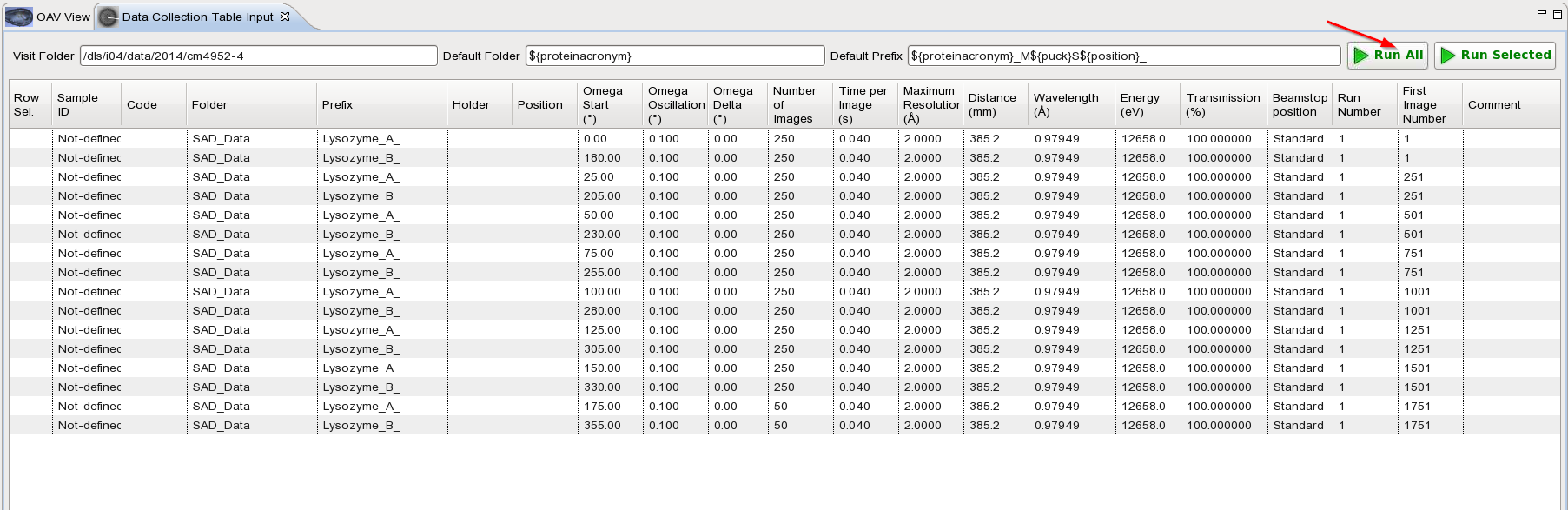
The results will appear as 2 distinct data sets: Lysozyme_A and Lysozyme_B. You will need to merge the data sets to process it later on.


  • Contact Us
  • About Diamond Light Source
  • Procurement
  • Supply Chain Transparency
  • Cookie Policy
  • Website Terms of Use
  • Privacy Notice

Diamond Light Source

Diamond Light Source is the UK's national synchrotron science facility, located at the Harwell Science and Innovation Campus in Oxfordshire.

Follow us on Facebook Follow us on Linkedin Follow us on Instagram Follow us on Bluesky

Diamond Light Source Ltd
Diamond House
Harwell Science & Innovation Campus
Didcot
Oxfordshire
OX11 0DE

See on Google Maps

Copyright © Diamond Light Source. Diamond Light Source® and the Diamond logo are registered trademarks of Diamond Light Source Ltd

Registered in England and Wales at Diamond House, Harwell Science and Innovation Campus, Didcot, Oxfordshire, OX11 0DE, United Kingdom. Company number: 4375679. VAT number: 287 461 957. Economic Operators Registration and Identification (EORI) number: GB287461957003.

feedback