eBIC (the Electron Bio-Imaging Centre)
eBIC took its first users in June 2015 and has since grown to be the largest national high-end cryoEM centre worldwide.
The centre provides scientists with state-of-the-art experimental equipment and expertise in the field of cryo-electron microscopy, for both single particle analysis and cryo-tomography. Designed for rapid, stable, high-resolution data collection on frozen-hydrated samples, eBIC offers Technology Track (high resolution data collection) and Signature Access (in-cell structural biology; EM tomography) via iNEXT DIscovery.
eBIC offers access to:
Single Particle Cryo-electron Microscopy
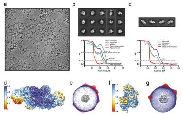
This technique requires the collection of a large number of movies from mono-disperse protein complexes or viruses such that their 3D structure can be determined. The Titan Krios microscopes are equipped with direct electron detectors and automated data collection software that allow a large number of movies to be collected from single particle samples. The type of single particles that can be imaged range from small protein complexes (150 kDa) to large viruses (2 MDa). For proteins smaller than 150 kDa the use of the Volta phase plate may be required.
Cryo-electron Tomography
Molecular
This technique can be used with single particle samples and is excellent at generating initial models for single particle analysis or for analysing repeating structures in larger pleomorphic objects. Tilt series are collected at areas of interest, which can then be aligned and reconstructed to generate 3D volumes. For thicker specimens zero-loss imaging is recommended.
Cellular
This technique is used to look at large pleomorphic objects such as vesicles, isolated organelles, bacteria, and intact eukaryotic cells. Tilt series are collected at areas of interest which can then be aligned and reconstructed to generate 3D volumes. For thicker specimens zero-loss imaging is recommended and a maximum sample thickness is of 0.5 um is advised.
Sample preparation by cryoFIB milling
The primary use of the Aquilos 2 cryo-FIB DualBeam is to generate lamellae for cryogenic TEM experiments (mainly tomography but also microED). Lamellae are thin sections of the target e.g. cells or crystals, that have been produced by milling away excess material in stages where the ion-beam current is reduced each time. This is done to avoid local heating whilst also increasing the milling rate. Typically, the lamellae are ~20° from the plane of the grid and are generally less than 300nm thick.
Dr Daniel Clare: [email protected]
Dr Yuriy Chaban: [email protected]
Dr Karen Davies: [email protected]

Diamond Light Source is the UK's national synchrotron science facility, located at the Harwell Science and Innovation Campus in Oxfordshire.
Copyright © 2022 Diamond Light Source
Diamond Light Source Ltd
Diamond House
Harwell Science & Innovation Campus
Didcot
Oxfordshire
OX11 0DE
Diamond Light Source® and the Diamond logo are registered trademarks of Diamond Light Source Ltd
Registered in England and Wales at Diamond House, Harwell Science and Innovation Campus, Didcot, Oxfordshire, OX11 0DE, United Kingdom. Company number: 4375679. VAT number: 287 461 957. Economic Operators Registration and Identification (EORI) number: GB287461957003.

 For Users