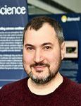
Principal Electron Microscopist for the electron Bio-Imaging Centre (eBIC)
Yuriy joined eBIC in March 2017 from the Imperial College London.
Email: yuriy.chaban@diamond.ac.ukTel: +44 (0) 1235 77 8207

Principal Electron Microscopist for the electron Bio-Imaging Centre (eBIC)
Yuriy joined eBIC in March 2017 from the Imperial College London.
Email: yuriy.chaban@diamond.ac.ukDr Yuriy Chaban graduated from Department of Genetics and Molecular Biology, Biological Faculty, Odesa II Mechnikov National University, Odesa, Ukraine in 1996. After graduation (1996-2000)
he worked as a Research Associate at the Department of Biochemistry at the same University, where he studied bioenergetics of cyanobacteria in the lab of Dr II Brown.
During his Ph.D. study (2000-2005) in the group of Prof. Egbert J Boekema at the Department of Biophysical Chemistry, University of Groningen, the Netherlands, Yuriy worked on structural characterisation of V-type ATPase and related enzymes. In 2005 Yuriy joined for his post-doctoral training, the lab of Dr Francisco J Asturias at The Scripps Research Institute, San Diego, USA, where he contributed to structural studies of RSC Chromatin Remodelling complex and Transcription Initiation Complexes.
In 2008 Yuriy moved to the UK to continue his post-doctoral training in the lab of Prof. Elena V Orlova at the Institute of Molecular and Structural Biology, Birkbeck College, London, UK. During his time at Birkbeck, Yuriy worked on the structures of SPP1 bacteriophage head-to tail interface and Bovine Papilloma Virus E1 helicase.
In 2012 he joined the lab of Prof. Dale B Wigley at the Institute of Cancer Research, London, UK, to work on structural characterisation of SWR1 chromatin remodelling complex. In 2013 he was promoted to staff scientist post. In 2015 Yuriy moved with the Prof Wigley lab to Imperial College London, UK. Here, in addition to SWR1 work, he contributed to structural studies of RecBCD helicase-nuclease complex.
Yuriy joined eBIC team at Diamond Light Source Ltd in 2017 as a cryo-EM scientist. In 2019 Yuriy was promoted to senior cryo-EM scientist post, and in January 2021 to Principal Electron Microscopist.
Cheng K, Wilkinson M, Chaban Y, Wigley DB A Conformational switch in response to Chi converts RecBCD from phage destruction to DNA repair., Nat Struct Mol Biol., 2020, 1, pp71-77.
Xiao J, Liu M, Qi Y, Chaban Y, Gao C, Pan B, Tian Y, Yu Z, Li J, Zhang P, Xu Y, Structural insight into the activation of ATM kinase., Cell Res., 2019, 8, pp683-685.
Willhoft O, Ghoneim M, Lin C-L, Chua EYD, Wilkinson M, Chaban Y, Ayala R, McCormack EA, Ocloo L ,Rueda DS, Wigley DB, Structure and dynamics of the yeast SWR1-nucleosome complex. Science, 2018, 362, eaat7716.
Lin CL, Chaban Y, Rees DM, McCormack EA, Ocloo L, Wigley DB, Functional characterization and architecture of recombinant yeast Swr1 histone exchange complex., Nucleic Acids Res., 2017, 45 (12) pp: 7249-7260.
Wilkinson M, Troman L, Ismah WWN, Chaban Y, Avison M, Dillingham M, Wigley D, Structural basis for the inhibition of RecBCD by Gam and its synergistic antibacterial effect with quinolones., eLife, 2016;5: e22963.
Wilkinson M, Chaban Y, Wigley DB, Mechanism for nuclease regulation in RecBCD., eLife, 2016;5:e18227.
Chaban Y, Lurz R, Brasiles S, Cornilleau C, Karreman M, Zinn-Justin S, Tavares P, Orlova EV., Structural rearrangements in the phage head-to-tail interface during assembly and infection., Proc Natl Acad Sci USA, 2015, pp:7009-7014.
Chaban Y, Stead J, Ryzhenkova K, Whelan F, Lamber EP, Antson A, Sanders CM, Orlova EV., Structural basis for DNA strand separation by a hexameric replicative helicase., Nucleic Acid Research, 2015, pp: 8551-63.
Hesketh EL, Parker-Manuel RP, Chaban Y, Satti R, Coverley D, Orlova EV, Chong JP., DNA induces conformational changes in a recombinant human minichromosome maintenance complex., J.Biol.Chem., 2015, pp:7973-7979.
Chaban Y, Boekema EJ, Dudkina NV., Structures of mitochondrial oxidative phosphorylation supercomplexes and mechanisms for their stabilisation., Biochim.Biophys.Acta., 2014, pp:418-426.
Cai G, Chaban YL, Imasaki T, Kovacs JA, Calero G, Penczek PA, Takagi Y, Asturias FJ. Interaction of the mediator head module with RNA polymerase II., Structure, 2012, pp:899-910.
Chittuluru JR, Chaban Y, Monnet-Saksouk J, Carrozza MJ, Sapountzi V, Selleck W, Huang J, Utley RT, Cramet M, Allard S, Cai G, Workman JL, Fried MG, Tan S, Côté J, Asturias FJ., Structure and nucleosome interaction of the yeast NuA4 and Piccolo-NuA4 histone acetyltransferase complexes., Nat. Struct .Mol .Biol., 2011, pp. 1196-203.
Okorokov AL, Chaban YL, Burgeev DV, Hodgkinson J, Mazin AV, Orlova EV., Structure of the hDmc1-ssDNA filaments reveals the principle of its architecture., PLoS One, 2010, 5(1):e8586.
Chaban Y, Ezeokonkwo C, Chung WH, Zhang F, Kornberg RD, Maier-Davis B, Lorch Y, Asturias FJ., Structure of a RSC-nucleosome complex and insight into chromatin remodeling., Nat .Struct .Mol. Biol., 2008, pp. 1272-7.
Chaban Y.L., Juliano S., Boekema E.J., Grüber G. Interaction between subunit C (Vma5p) of the yeast vacuolar ATPase and the stalk of the C-depleted V(1) ATPase from Manduca sexta midgut., Biochim Biophys. Acta, 2005, 1708, pp. 196-200.
Chaban Y.L., Coskun Ü., Keegstra W., Oostergetel G., Boekema E.J. and Grüber G. Structural characterization of an ATPase active F1-/V1-ATPase (alpha3beta3EG) hybrid complex., J Biol. Chem., 2004, 279, pp. 47866-47870.
Coskun Ü., Chaban Y.L., Lingl A., Müller V., Keegstra W., Boekema E.J. and Grüber G. Structure and subunit arrangement of the A-type ATP synthase complex from the archaea Methanococcus jannaschii vizualized by electron microscopy., J Biol. Chem., 2004, 279, pp. 38644-38648.
Chaban Y., Ubbink-Kok T., Keegstra W., Lolkema J.S. and Boekema E.J. Composition of the central stalk of the Na+-pumping V-ATPase from Caloramator fervidus, EMBO reports., 2002., 3, (10), pp. 982-987.
Brown I.I., Ishmukhametov R.R., Karakis S.G., Pogorelov D.I., Chaban Y.L., Adaptation characters of cyanobacteria from the different hydrocenoses to alkaline conditions.- Ecologiya Morya (Ecology of the Sea), 2001, 55, pp. 53-58 (Russ.).
Chaban Y.L., Ishmukhametov R.R. Photosynthesis of the cyanobacterium Gloeobacter violaceus reveals dependence from the cation composition.- Wistnyk ODU (Bulletin of Odessa State University), 2000, 5, 1, pp. 47-53 (Ukr.) .
Brown I. I., Chaban Y., Ishmukhametov R., Lovenchuk I., Karakis S., Pogorelov D. (1999) The peculiarities of bioenergetics coupling in cyanobacteria under low Δμ H+ and ΔμNa+ in Marine cyanobacteria, 1999. Charpy L & Larkum AWD (eds.) Bulletin de l'Institut Oceanographique, Monaco, special issue, pp. 229 - 235.
Diamond Light Source is the UK's national synchrotron science facility, located at the Harwell Science and Innovation Campus in Oxfordshire.
Copyright © 2022 Diamond Light Source
Diamond Light Source Ltd
Diamond House
Harwell Science & Innovation Campus
Didcot
Oxfordshire
OX11 0DE
Diamond Light Source® and the Diamond logo are registered trademarks of Diamond Light Source Ltd
Registered in England and Wales at Diamond House, Harwell Science and Innovation Campus, Didcot, Oxfordshire, OX11 0DE, United Kingdom. Company number: 4375679. VAT number: 287 461 957. Economic Operators Registration and Identification (EORI) number: GB287461957003.