Instruments by Science Group
B22 Contact
Beamline Phone Number:
+44 (0) 1235 778684
Principal Beamline Scientist:
Dr Gianfelice Cinque
Tel: +44 (0) 1235 778410
E-mail: [email protected]
Science Group Leader
Robert Rambo
Email: [email protected]
Tel: +44 (0)1235 56 7675
B22 Multimode InfraRed Imaging And Microspectroscopy
Status: Operational
Beamsize: 15µm to 3 µm by microFTIR; resolution 10 to 100 nm by nanoIR (s-SNOM & AFM IR)
Detector: MCT, FPA, DLaTGS and Bolometer
Wavelength: 1 mm - 1 µm (10,000 to 10 cm-1)
Energy: 1 meV - 1 eV
Detector: MCT, FPA, DLaTGS and Bolometer
Wavelength: 1 mm - 1 µm (10,000 to 10 cm-1)
Energy: 1 meV - 1 eV
Operating principle
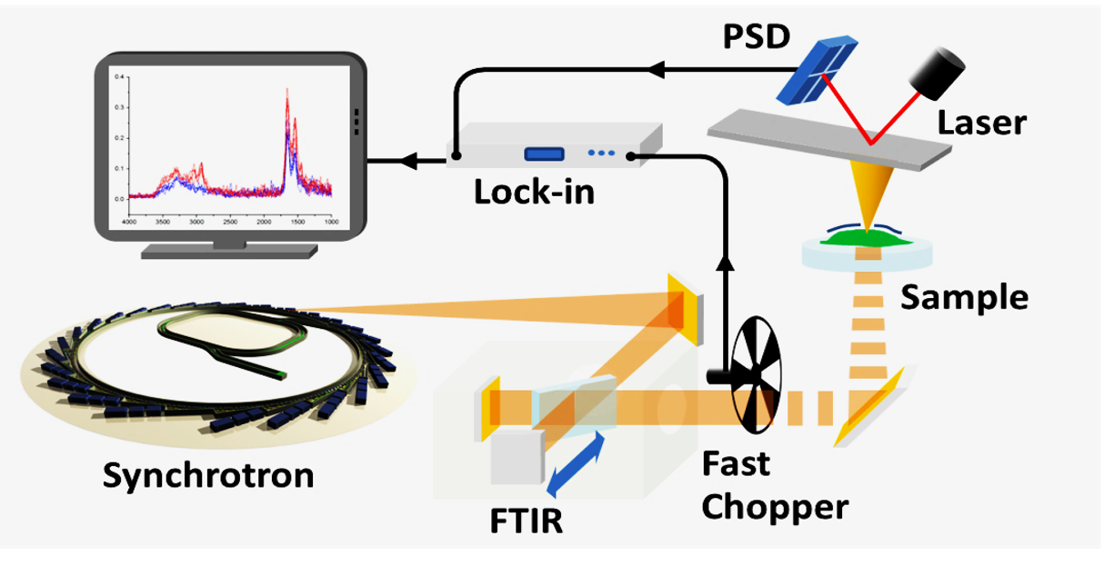
- Setup for synchrotron-based PTE spectroscopy
The working principle of AFM-IR spectroscopy is based on the unification of the mechanical sensitivity of AFM detection - enhanced in resonance - with the chemical sensitivity of IR spectroscopy:
- The sample is scanned using an AFM tip in contact, allowing for nanoscale characterization of mechanical properties and sample topography
- A pulsed IR source illuminates the sample surface: for Synchrotron radiation, a fast light chopper is used
- Upon absorption of IR light, the sample undergoes photo-thermal expansion, typically on the picometer scale
- The AFM tip can measure the thermal expansion after mechanical enhancement of the cantilever deflection working in resonance with the light chopper modulation
- The detection occurs as a mechanical signal using a symmetric FTIR interferometer
- The system can be operated in both top and bottom illumination
Magnetic levitation in vacuum light chopper
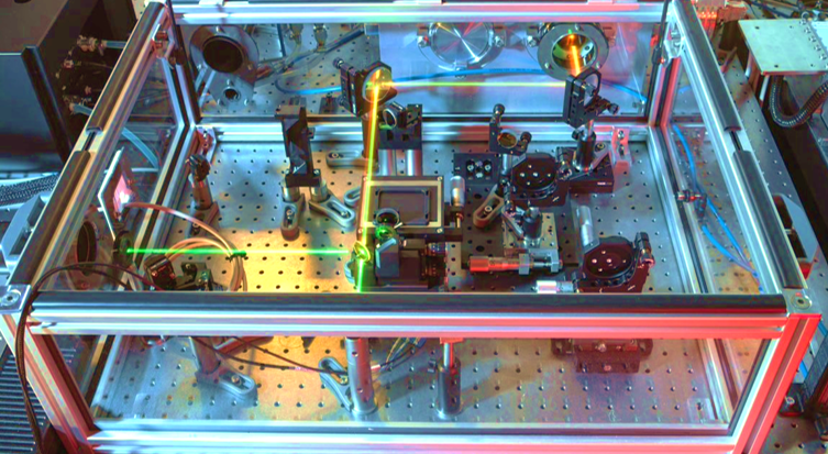
- The external beampath, coupling light from the chopper (top right) to the neaScope (bottom left) with the interferometer stage in the centre
In the present setup the photothermal expansion is detected using AFM in a resonance-enhanced contact mode. Resonance enhancement can improve the signal by bringing the modulation frequency of the light in resonance with the frequency of the cantilever. In order to do so, for a quasi-continuous source, such as SRIR, a fast chopper is required.
Together with Celeroton, Switzerland, a bespoke magnetic levitating vacuum chopper system was developed by B22 beamline staff at DLS. This allows to reach modulation frequencies of up to 200 kHz, corresponding to the second contact resonance of widely used AFM tips.
A detailed description of the chopper setup has been published in:
Some key features
- Non surface sensitive, suitable for micron-thick samples (e.g. cells)
- Transmission illumination available
- Nano-spectra truly corresponding to vibrational absorption spectra
- Entire IR spectral range accesible, including THz
- Spatial resolution on the order of 100s nm, dictated by sample/substrate thermal diffusivity
- Relies on thermo-mechanical expansion of the sample.
The method is most suitable for biological samples (e.g. mammalian or plant cells), as the whole
mid-IR range can be studied and the full thickness of samples on the order of 1 micron is probed.
Selected relevant publications
Publications on the technique:
- Dazzi, Alexandre, et al. "AFM–IR: combining atomic force microscopy and infrared spectroscopy for nanoscale chemical characterization." Applied spectroscopy 66.12 (2012): 1365-1384.
- Dazzi, Alexandre, and Craig B. Prater. "AFM-IR: Technology and applications in nanoscale infrared spectroscopy and chemical imaging." Chemical reviews 117.7 (2017): 5146-5173.
- Mathurin, Jeremie, et al. "Photothermal AFM-IR spectroscopy and imaging: Status, challenges, and trends." Journal of Applied Physics 131.1 (2022).
Publications on projects at B22 (related to the previous setup):
- Synchrotron photothermal IR nanospectroscopy of macrophages drug-induced phospholipidosis- Ka Lung Andrew Chan, Ioannis Lekkas, Mark D. Frogley, Gianfelice Cinque, Ali Altharawi, Gianluca Bello, Lea Ann Dailey - Analytical Chemistry, May 2020 DOI: 10.1021/acs.analchem.9b05759
- Performances for broadband synchrotron photothermal infrared nano-spectroscopy at Diamond Light Source - Mark D. Frogley, Ioannis Lekkas, Chris S. Kelley, Gianfelice Cinque - Infrared Physics & Technology, Feb 2020 DOI: 10.1016/j.infrared.2020.103238
- Synchrotron infrared near-field spectroscopy in photothermal mode - Chris S. Kelley, Mark D. Frogley, Ann Fitzpatrick, Katia Wehbe, Paul Donaldson, Gianfelice Cinque - Spectroscopy Europe, 28
- World first for Diamond in synchrotron-based IR photothermal nanospectroscopy-Gianfelice Cinque, Chris S. Kelley, Mark D. Frogley, Jacob Filik, Katia Wehbe, Ann Fitzpatrick, Paul M. Donaldson - Synchrotron Radiation News, 29, Aug 2016 DOI: 10.1080/08940886.2016.1198675

Diamond Light Source is the UK's national synchrotron science facility, located at the Harwell Science and Innovation Campus in Oxfordshire.
Diamond Light Source Ltd
Diamond House
Harwell Science & Innovation Campus
Didcot
Oxfordshire
OX11 0DE
Copyright © Diamond Light Source. Diamond Light Source® and the Diamond logo are registered trademarks of Diamond Light Source Ltd
Registered in England and Wales at Diamond House, Harwell Science and Innovation Campus, Didcot, Oxfordshire, OX11 0DE, United Kingdom. Company number: 4375679. VAT number: 287 461 957. Economic Operators Registration and Identification (EORI) number: GB287461957003.