Grid Scan
The diffraction grid scan is a particularly useful function. It can help with:
- Locating small crystals in the loop
- Locating crystals in opaque mother liquors (e.g. LCP)
- Identifying the optimal diffraction from a large crystal
- X-ray centring on a crystal
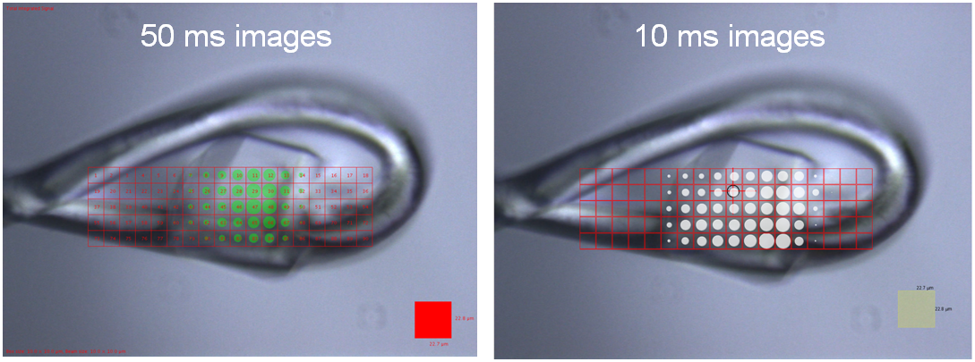
Ideally, your initial grid scan will have the loop face on to the X-ray beam. You can then identify the best part of the crystal. Centre on the best part of the crystal and rotate the sample by 90° (not in in-situ mode). You can then draw a single column grid to assess the diffraction from this orientation. By carrying out two grid scans like this you can easily centre on the best part of the crystal.
Running a Grid Scan
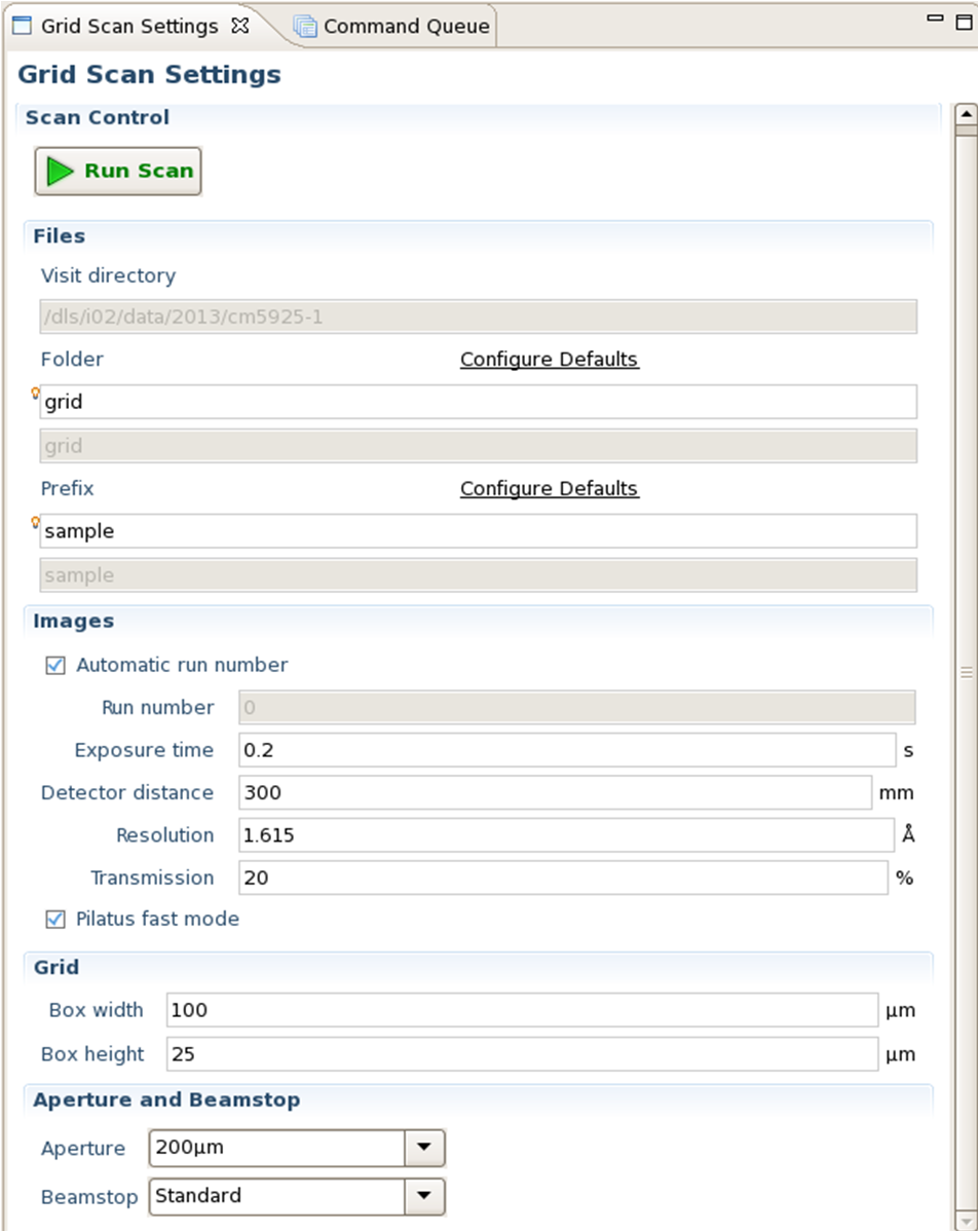
The diffraction grid and scan settings are accessed from the 'Grid Scan' perspective.
The grid to test can be drawn on the sample in the OAV image on the left side of the perspective. This is done by left clicking dragging over the area of interest on the screen. To erase a previous grid and draw another simply carry out this process starting beyond the edge of the existing grid.
To move the grid, left click and drag with the pointer on the exisiting grid.
Instructions for drawing and manipulating the grid are given under the OAV image of the crystal on the left side of the perspective.
On the right panel of the perspective you can set the parameters for the data collection within the grid scan. Please note that it is worth using low transmission for grid scans to minimise the radiation damage to your sample.
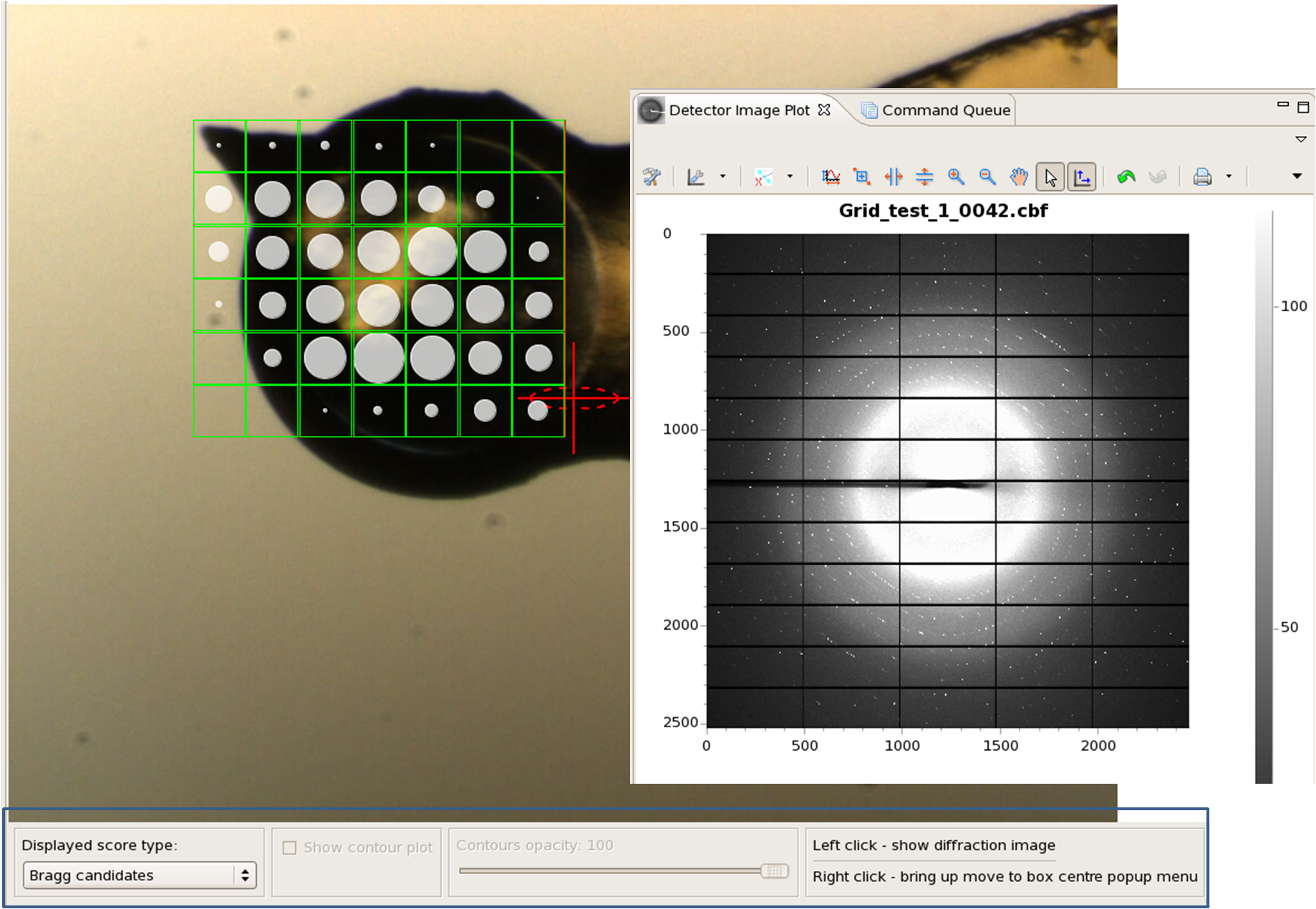
Grid Scan Results
Grid scan results are displayed in the 'Grid Scan Results' perspective.
The grid turns green as the data for each box is collected. By default images are ranked by the size of the circle in the box which represents the number of Bragg candidates in the collected data for that box. This criteria can be changed using the drop down menu on the bottom left hand side of the OAV image (blue box).
As data are collected the images are displayed in the Detector Image Plot which opens by default in the right hand panel of GDA. To view the diffraction image of any box in the grid scan in this viewer, simply left click in the box of interest.
Having decided where to centre the crystal, simply right click on the box of interest and chose 'move to box centre'. This will align that box with the X-rays ready for data collection.