
Principal Electron Microscopist for the electron Bio-Imaging Centre (eBIC)
Vojtech joined eBIC in April 2026.
Email: [email protected]Tel: +44 (0) 1235 77 8131
Principal Electron Microscopist for the electron Bio-Imaging Centre (eBIC)
Vojtech joined eBIC in April 2026.
Email: [email protected]Vojta Pražák is a structural cell biologist developing and applying advanced cryogenic light and electron microscopy to study host-pathogen interactions in situ. His work focuses on resolving molecular mechanisms at the interface between pathogens and their hosts, integrating data across scales from single proteins to cells and whole tissues. His current work is centred on understanding the molecular basis of interactions between filamentous pathogens (fungi and oomycetes) and their hosts.
He develops and applies methodologies that bring advanced cryogenic imaging into routine use, linking high-resolution structural methods with established experimental systems. In close collaboration with Prof Rainer Kaufmann (University of Hamburg), Prof Lindsay Baker (University of Oxford) and Prof Kay Grünewald (University of Hamburg), he has contributed to the development of super-resolution cryo-correlative light and electron microscopy (cryoCLEM). This technique bridges cell and structural biology by utilising conventional fluorescent protein tags to localise specific complexes within high-resolution electron cryo-tomography data. In parallel, he has developed computational tools for cryoET for structural determination using subvolume averaging in complex cellular environments.
Vojta values existing partnerships and actively seeks to develop new ones with collaborators who value openness, rigour, and mutual respect. In addition to those listed above, he maintains established collaborations with Prof Emmanuelle Quemin (I2BC), Dr Emma Silvester (University of Oxford), Dr Emily Machala (University of Oxford), Prof Christiane Riedel (Université de Lyon), Prof Pieter van West (University of Aberdeen), Prof Tolga Bozkurt (Imperial College London), Prof Paul Birch (University of Dundee), and Prof Ronelle Roth (University of Oxford). Above all, Vojta’s work is guided by a commitment to advancing our understanding of filamentous pathogens and conducting research in a manner that is kind and considerate of people and the wider living world.
Corroyer-Dulmont, S., Labarde, A., Pražák, V., Godinho, L., Masson, C., Legrand, P., Grünewald, K., Tavares, P., Quemin, E.R.J. Subcellular reorganization upon phage infection reveals stepwise assembly of viral particles from membrane-associated precursors. Nature Communications (2026) https://doi.org/ 10.1038/s41467-026-71181-w
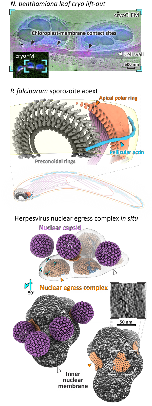
Yuen, ELH., Savage, Z., Pražák, V., Adamkova, V., King,F., Vuolo, C., Ibrahim, T., Jenkins, S., Wang, Y., and Zhou, Y. et al., 2025. Membrane Contact Sites Between Chloroplasts and Pathogen Interface Underpin Plant Focal Immune Responses. The Plant Cell (2025). https://doi.org/10.1093/plcell/koaf214
Pražák, V., Vasishtan, D., Grünewald, K., Douglas, R.G. and Ferreira, J.L. Molecular architecture of glideosome and nuclear F-actin in Plasmodium falciparum. EMBO Rep (2025). https://doi.org/10.1038/s44319-025-00415-7
Hofstadter, W. A., Cook, K. C., Tsopurashvili, E., Gebauer, R., Pražák, V., Machala, E. A., Park, J. W., Grünewald, K. et al. Infection-induced peripheral mitochondria fission drives ER encapsulations and inter-mitochondria contacts that rescue bioenergetics. Nature Communications (2024). https://doi.org/10.1038/s41467-024-51680-4
Pražák, V.*, Mironova, Y.*, Vasishtan, D., Hagen, C., Laugks, U., Jensen, Y., Sanders, S. et al. Molecular plasticity of herpesvirus nuclear egress analysed in situ. Nature Microbiology (2024). https://doi.org/10.1038/s41564-024-01716-8
Liu, J., Corroyer-Dulmont, S., Pražák, V., Khusainov, I., Bahrami, K., Welsch, S., Vasishtan, D. et al. The palisade layer of the poxvirus core is composed of flexible A10 trimers. Nat Struct Mol Biol (2024). https://doi.org/10.1038/s41594-024-01218-5
Ferreira, J. L.*, Pražák, V.*, Vasishtan, D., Siggel, M., Hentzschel, F., Binder, A. M., Pietsch, E. et al. Variable microtubule architecture in the malaria parasite. Nature Communications (2023). https://doi.org/10.1038/s41467-023-36627-5
Villalta, A., Schmitt, A., Estrozi, L. F., Quemin, E. R. J., Alempic, J.-M., Lartigue, A., Pražák, V. et al. The giant mimivirus 1.2 Mb genome is elegantly organized into a 30-nm diameter helical protein shield. eLife (2022). https://doi.org/10.7554/eLife.77607
Silvester, E., Vollmer, B., Pražák, V., Vasishtan, D., Machala, E. A., Whittle, C., Black, S. et al. DNA origami signposts for identifying proteins on cell membranes by electron cryotomography. Cell (2021). https://doi.org/10.1016%2Fj.cell.2021.01.033
Pražák, V., Grünewald, K. & Kaufmann, R. Correlative super-resolution fluorescence and electron cryo-microscopy based on cryo-SOFI Methods in Cell Biology (2021). https://doi.org/10.1016/bs.mcb.2020.10.021
Vollmer, B., Pražák, V., Vasishtan, D., Jefferys, E. E., Hernandez-Duran, A., Vallbracht, M., Klupp, B. G. et al. The prefusion structure of herpes simplex virus glycoprotein B. Science Advances (2020). https://doi.org/10.1126%2Fsciadv.abc1726
Quemin, E. R. J., Machala, E. A., Vollmer, B., Pražák, V., Vasishtan, D., Rosch, R., Grange, M. et al. Cellular Electron Cryo-Tomography to Study Virus-Host Interactions. Annual review of virology (2020). https://doi.org/10.1146/annurev-virology-021920-115935
Ni, T., Jiao, F., Yu, X., Aden, S., Ginger, L., Williams, S. I., Bai, F., Pražák, V. et al. Structure and mechanism of bactericidal mammalian perforin-2, an ancient agent of innate immunity. Science Advances (2020). https://doi.org/10.1126/sciadv.aax8286
Riedel, C., Vasishtan, D., Pražák, V., Ghanem, A., Conzelmann, K. K. & Rümenapf, T. Cryo EM structure of the rabies virus ribonucleoprotein complex. Scientific reports (2019). https://doi.org/10.1038/s41598-019-46126-7
Moser*, F., Pražák*, V., Mordhorst, V., Andrade, D. M., Baker, L. A., Hagen, C., Grünewald, K. & Kaufmann, R. Cryo-SOFI enabling low-dose super-resolution correlative light and electron cryo-microscopy. Proceedings of the National Academy of Sciences of the United States of America (2019). https://doi.org/10.1073/pnas.1810690116
Böttcher, B., Pražák, V., Rasmussen, A., Black, S. S. & Rasmussen, T. The Structure of YnaI Implies Structural and Mechanistic Conservation in the MscS Family of Mechanosensitive Channels. Structure (2015). https://doi.org/10.1016/j.str.2015.06.023

Diamond Light Source is the UK's national synchrotron science facility, located at the Harwell Science and Innovation Campus in Oxfordshire.
Diamond Light Source Ltd
Diamond House
Harwell Science & Innovation Campus
Didcot
Oxfordshire
OX11 0DE
Copyright © Diamond Light Source. Diamond Light Source® and the Diamond logo are registered trademarks of Diamond Light Source Ltd
Registered in England and Wales at Diamond House, Harwell Science and Innovation Campus, Didcot, Oxfordshire, OX11 0DE, United Kingdom. Company number: 4375679. VAT number: 287 461 957. Economic Operators Registration and Identification (EORI) number: GB287461957003.