• Diamond Home page
  • About
  • For Users
  • Public
  • Industry
  • Instruments
  • Careers
  • More Show more menu items
Search

About

  • About Diamond
  • About Synchrotrons
  • News and Features
  • Events
  • Contact Us
  • FAQ

For Users

  • Apply for beamtime
  • User guide
  • Diamond Users Commitee
  • FAQ
  • User Administration System
  • iSpyB

Industry

  • Techniques Available
  • Industry Research
  • Industry Case Studies
  • News
  • Meet the Industry Team

Science

  • Research
  • Computing
  • The Machine
  • Publications
  • Research Expertise
  • Membrane Protein Laboratory
  • Additional Facilities
  • Publications Database

Instruments

  • Biological Cryo-Imaging
  • Crystallography
  • Imaging and Microscopy
  • Macromolecular Crystallography
  • Magnetic Materials
  • Soft Condensed Matter
  • Spectroscopy
  • Structures and Surfaces

Diamond-II

  • Science
  • Machine
  • Beamlines
  • Software, Control and Computing
  • Infrastructure

Public

  • Public open days
  • School visits
  • Partner with Diamond
  • Explore Diamond

Procurement

  • Non-OJEU Tender Notices
  • OJEU PINs
  • OJEU Tender Notices
  • Registration Form

Careers

  • Vacancies
  • Info for applicants
  • Company Benefits
  • Apprenticeships
  • PhD Studentships
  • Work Placement

Main Content

Structures and Surfaces
Sub-navigation
  • Structures and Surfaces
  • I05
  • B07
  • I07
  • I09

In This Section

Sub Navigation
  • Beamline Guide
    • Optical Overview
    • B07-B: NEXAFS / UHV XPS
    • B07-C: AP-XPS
      • Endstation Setup
        • Sample Holders
        • Gas Rig
        • Detectors and Analysers
        • Cells at B07-C
      • Beamline/Analyser performance
    • Sample Preparation
    • Support laboratory
  • Techniques and Instrumentation
  • Scientific Applications
  • Rapid Access
  • Useful links
  • Manuals
  • Science Highlights and News
  • Working Group
  • Staff
  • FAQ
  • Collaborations
  • Publications
  • Accessibility Information

Instruments by Science Group

Macromolecular
Crystallography
Soft Condensed
Matter
Imaging and
Microscopy
Biological
Cryo-Imaging
Magnetic
Materials
Structures and
Surfaces
Crystallography
Spectroscopy

B07 Contact

Principal Beamline Scientist:
Georg Held

Tel: +44 (0) 1235 778480
E-mail: [email protected]

More

Science Group Leader

Cephise Cacho

Email: [email protected]
Tel: +44 (0)1235 778290

B07 VERSOX: Versatile Soft X-ray

Status: Operational

Wavelength: 24.8 - 0.44 nm
Energy: 50 - 2800 eV

XAS: X-ray Absorption Spectroscopy XPS: X-ray Photoelectron Spectroscopy NEXAFS: Near Edge X-ray Absorption Fine Structures NAP-XPS: Near Ambient Pressure XPS Spectroscopy
  1. Instruments
  2. Structures and Surfaces
  3. B07
  4. Beamline Guide
  5. B07-C: AP-XPS
  6. Endstation Setup
  7. Detectors and Analysers

Detectors and Analysers

B07-C Ambient Pressure XPS

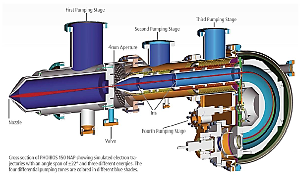
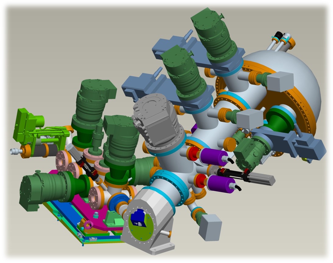
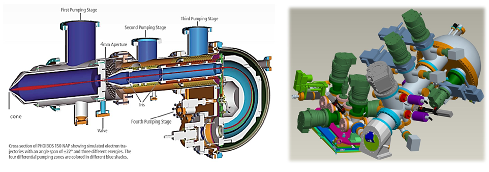
PHOIBOS 150 NAP Analyser

The electron energy analyser is a "PHOIBOS 150 NAP" hemispherical analyser supplied by SPECS, Berlin, Germany (left image). The analyser axis is at an angle of 60.1° with respect to the beam and tilted 30° with respect to the horizontal, i.e. close to the magic angle with respect to the polarization vector (right image). The entire assembly of interface ange and analyser is mounted on a "sledge" which can be moved parallel to the analyser axis. This way the cone-to-beam/sample distance can be changed without having to re-align the endstation, once the beam and the analyser axis have been adjusted such that they intersect in one point. The typical working distance between cone and sample is 0.2 - 0.3 mm. Compensation of residual magnetic fields is achieved via three pairs of Helmholtz coils, are individually controlled as a function of electron energy through the control unit of the electron energy analyser.

Cross secrion of PHOIBOS

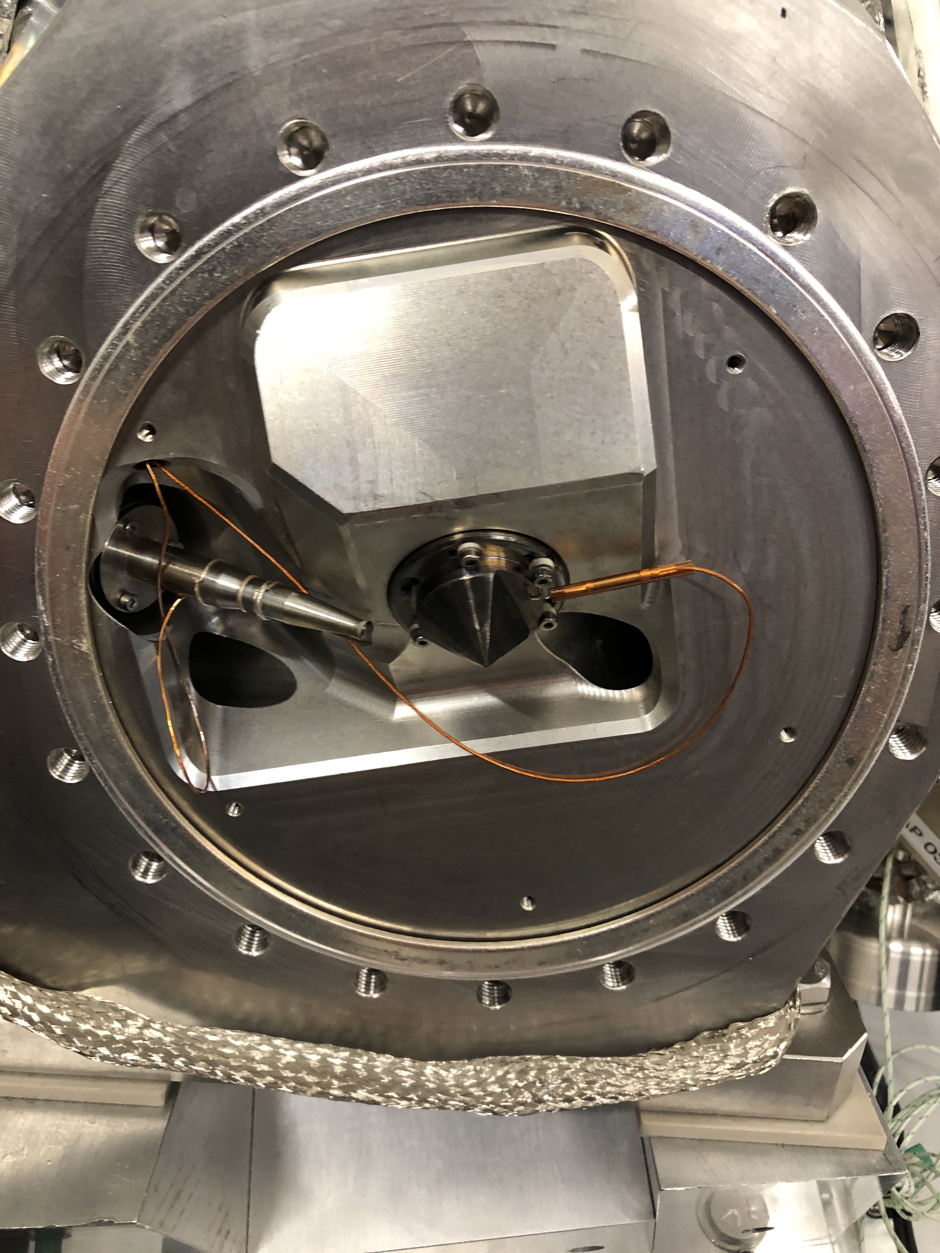
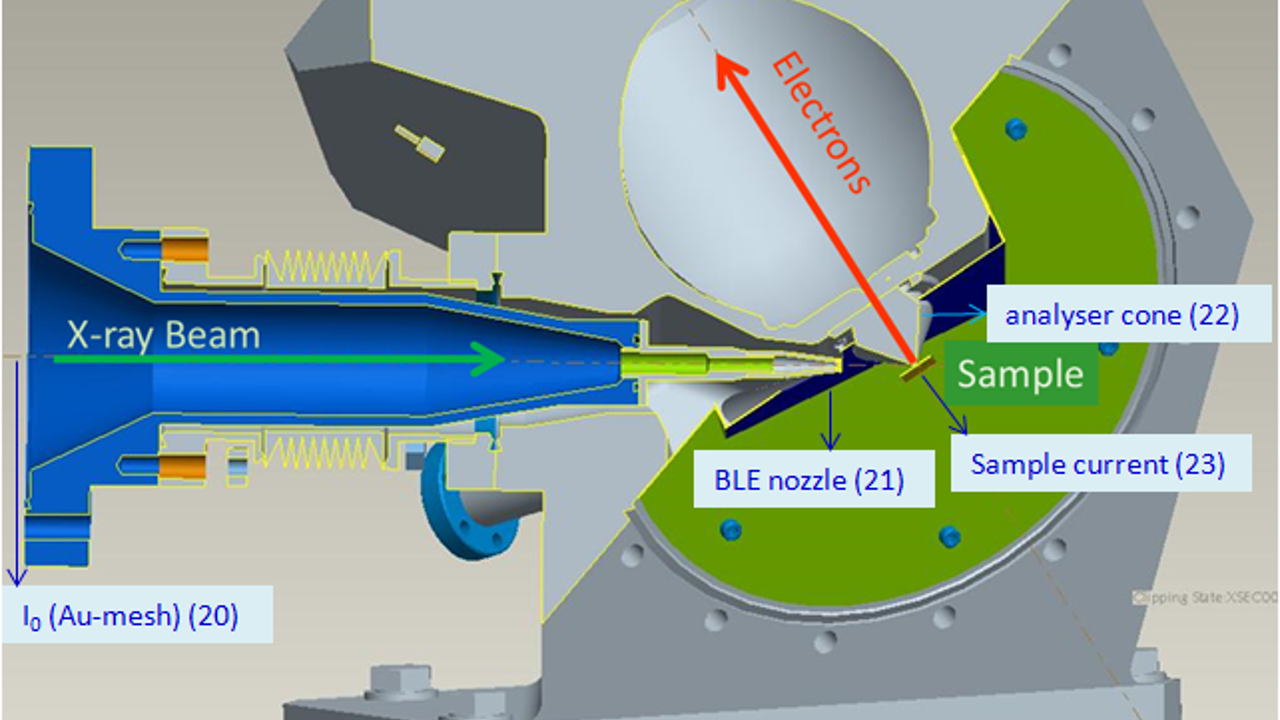
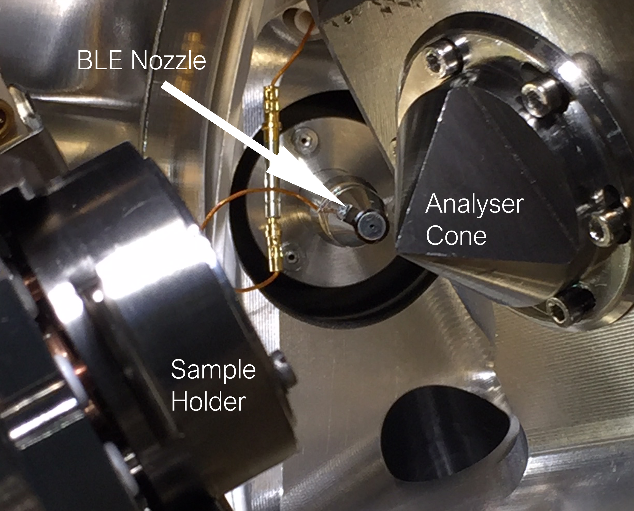
Total Electron Yield – Drain current for NEXAFS


The entry cone of the analyser's pre-lens has an aperture of 0.3mm and is isolated from ground to allow collection of TEY NEXAFS by measuring the drain current through a Stanford Research SR 570 current amplifier (image below). In total, four currents can be recorded simultaneously:

Signal Current Amplifier (old GDA name) Current Amplifier (GDA name)
 I0 (Au mesh)  ca20 ca31c
 BLE nozzle  ca21 ca32c
 analyser cone  ca22 ca33c
 sample drain current  ca23 ca34c


test Detector layout  cone

Current Amplifier Squemes: left, centre and right


  • Contact Us
  • About Diamond Light Source
  • Procurement
  • Supply Chain Transparency
  • Cookie Policy
  • Website Terms of Use
  • Privacy Notice

Diamond Light Source

Diamond Light Source is the UK's national synchrotron science facility, located at the Harwell Science and Innovation Campus in Oxfordshire.

Follow us on Facebook Follow us on Linkedin Follow us on Instagram Follow us on Bluesky

Diamond Light Source Ltd
Diamond House
Harwell Science & Innovation Campus
Didcot
Oxfordshire
OX11 0DE

See on Google Maps

Copyright © Diamond Light Source. Diamond Light Source® and the Diamond logo are registered trademarks of Diamond Light Source Ltd

Registered in England and Wales at Diamond House, Harwell Science and Innovation Campus, Didcot, Oxfordshire, OX11 0DE, United Kingdom. Company number: 4375679. VAT number: 287 461 957. Economic Operators Registration and Identification (EORI) number: GB287461957003.

feedback