Instruments by Science Group
Call for Proposals AP39
AP39 covers the period April 2026 - September 2026.
Proposals deadline: 1st Oct 2025 at 17:00hrs UTC/GMT
For the latest Equipment & Capabilities update check the individual beamline websites.
News Story March 2024
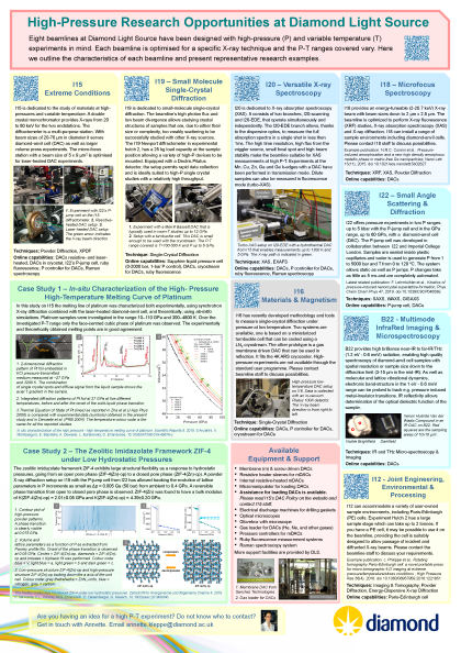
High-Pressure Research Opportunities at DLS
High-Pressure Research Day
9th Jan 2020 @ the Hilton Garden Inn, Abingdon - internal event
For more information email [email protected]
Useful Links
- EHPRG The European High Pressure Research Group
- AIRAPT International Association for the Advancement of High Pressure Science and Technology
- IUCr High Pressure International Union of Crystallography Commission on High Pressure
- CSEC Centre for Science at Extreme Conditions
SAXS
Small Angle X-ray Scattering (SAXS) is a technique particularly suited for the structural investigation of partially ordered materials, usually applied to condensed matter systems. SAXS provides information about larger scale bulk microstructure within a sample, usually in the size range from approximately ~1nm up to around 300nm.
X-ray Powder Diffraction
Powder diffraction is used to examine small, weakly interacting crystals in random orientations. Many materials exist as powders or in polycrystalline form, including ceramics, metals, superconductor oxides, pharmaceuticals, geochemicals, zeolites and related porous solids, all of which can be studied with powder diffraction. The technique can be used to study polycrystalline materials such as metals and alloys.
EXAFS
Extended X-ray Absorption Fine Structure (EXAFS) is a member of the XAS family. The incident energy is scanned in order to measure the absorption coefficient. EXAFS is elemental specific and is used to determine the oxidation state as well as the first neighbours of the absorbing atom.
Single-Crystal Diffraction
Small molecule single-crystal diffraction is the most widely used technique for obtaining full three-dimensional structural information of solid-state crystalline materials. This allows characterisation of, for example, molecular packing, guests in a framework structure, the nature of intra- and intermolecular interactions, molecular conformation and static and dynamic disorder. Environmental cells can be used to probe materials under a range of non-ambient conditions.
Infrared
IR spectroscopy is a widely used and versatile method for analysis at the molecular scale. IR Microspectroscopy allows not only molecular identification but also spatial resolution, which is necessary to the understanding of the physical-chemical properties of the vast range of materials and surfaces that are organised on a microstructural level. These include biomedical samples like tissue or single cells that can be mapped in plots showing molecular composition versus position by IR imaging technique.
XPDF
X-ray atomic pair distribution function (PDF) is an X-ray scattering technique that can be used to study the local structure of materials at the atomic scale. The technique requires scattering data to be collected to very high scattering angles using high energy powder diffraction coupled with Fourier transform to produce the PDF data.
EDXD
Instead of using X-rays of single energy, which is like using light of a single wavelength or colour, energy-dispersive diffraction (EDXD) uses a multi-wavelength "white beam" of X-rays from the synchrotron. The white beam is diffracted by the sample, producing a spectrum with characteristic intensity peaks at specific photon energies. The position of these peaks provides information about the crystal structure of the sample. Shifts in peak position are used to measure internal strains. By scanning the sample through the beam, a 2D or 3D map of strains in the sample can be produced.
Tomography
X-ray tomography is the construction of a three dimensional image from two dimensional projections taken at different orientations (usually with phase contrast or absorption contrast imaging). The tuneability of synchrotron X-rays make it possible to provide increased contrast images, and the coherent nature and high intensity of synchrotron X-rays have led to significant developments in this field, particularly in phase tomography which in the past has required extremely complex instrumentation.
XAS
A range of X-ray Absorption Spectroscopy techniques are available at Diamond, including X-ray Absorption Near-Edge Structure (XANES), Extended X-ray Absorption Fine Structure (EXAFS), Resonant Inelastic X-ray Scattering (RIXS) and X-ray Emission Spectroscopy (XES).
At characteristic wavelengths the X-ray absorption of an element changes dramatically, these are called absorption edges. Near the absorption edge, the spectra may contain fine structure that reveals the electronic and geometrical environment of the absorbing atom. This technique is XANES; further from the edge EXAFS reveals the local atomic environment to the element. Both can be used to can follow reactions on timescales down to the millisecond.
XRF Imaging
X-ray Fluorescence (XRF) occurs when the inner shell electrons of atoms in the sample get excited by the incident X-ray photons (synchrotron beam) and subsequently release X-ray photon when the system relaxes, that is when the electrons transition from the higher energy levels of the atom to the vacant inner shell. The beauty of this process is that each secondary X-ray photon (sometimes called characteristic radiation) emitted from the sample has a specific energy which is a fingerprint of the atom from which it has originated.
GISAXS: Grazing Incidence SAXS
GISAXS is a member of the SAXS technique family and the small-angle analogue of GIWAXS. It is a surface technique used in the characterisation of nanoscale surfaces and thin films. Most commonly GISAXS is used to determine, on a nanoscale, the self-assembly and self-organisation of thin films.

Diamond Light Source is the UK's national synchrotron science facility, located at the Harwell Science and Innovation Campus in Oxfordshire.
Diamond Light Source Ltd
Diamond House
Harwell Science & Innovation Campus
Didcot
Oxfordshire
OX11 0DE
Copyright © Diamond Light Source. Diamond Light Source® and the Diamond logo are registered trademarks of Diamond Light Source Ltd
Registered in England and Wales at Diamond House, Harwell Science and Innovation Campus, Didcot, Oxfordshire, OX11 0DE, United Kingdom. Company number: 4375679. VAT number: 287 461 957. Economic Operators Registration and Identification (EORI) number: GB287461957003.