My Days at Diamond - Gwyndaf Evans
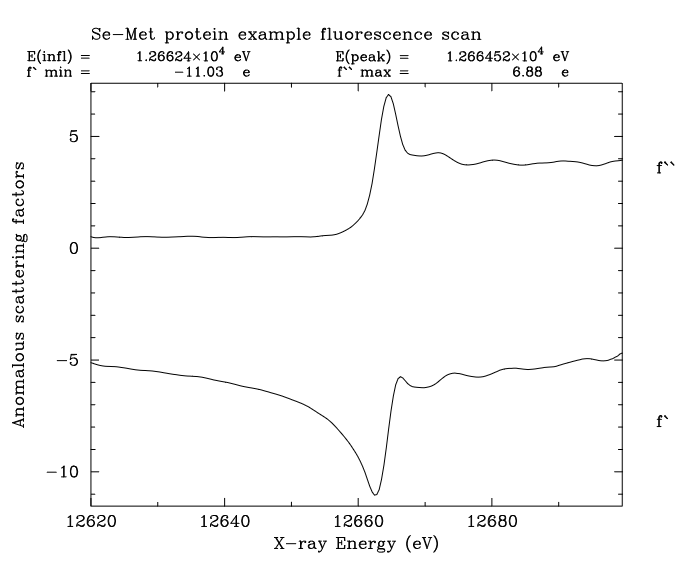
- Figure 1: An output from CHOOCH, taken from the publication.
Synchrotrons and CHOOCH
I began at Diamond on 6th January 2004. I moved there from Cambridge where I had worked for six years on data analysis methods and programming with Gerard Bricogne, first at the LMB and then at Global Phasing. There I became involved with CCP4, through Phil Evans and Andrew Leslie, and this link continued all the way to my retirement earlier this year. Before Cambridge, I had worked only at synchrotrons since 1989: EMBL-Hamburg and then the APS.
It was in Hamburg in the early 90s that I had written CHOOCH, a program for determining anomalous scattering factors from X-ray fluorescence spectra. At that time the method of multiwavelength anomalous diffraction (MAD) as popularised by Wayne Hendrickson and colleagues1 was growing and my PhD in Hamburg was focused around developing tools to allow beamlines to accurately measure MAD data: CHOOCH (Figure 1) was part of that work and eventually became part of the CCP4 suite. Those years in Hamburg laid the ground for my research interest which was the interface between the measurement and analysis of diffraction data in MX.
From Cambridge, I had been recruited to Diamond by Liz Duke, then MX PBS, and Dame Louise Johnson, Diamond’s first Life Science Director, to work with Liz on the three Phase 1 MX beamlines I02, I03 and I04. My initial focus was on the end-stations but within my first few months, with the Phase 2 beamline projects already underway, the I24 microfocus PBS job was being advertised. I applied and started in that role in the latter half of 2004. Given my previous experience, my interest in both the synchrotron instruments and the analysis of MX data, I was keen to bring these strands together as much as possible to benefit the design of the instruments and the software to get the most out of user samples.
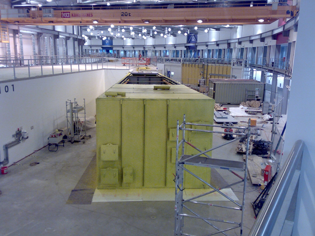
- Figure 2: The I24 hutch during construction in 2007
I24 and the MPL
In the 00s there was a major push to solve membrane protein structures and So Iwata, a major force in membrane protein crystallography joined Diamond in 2005 (I think) on a joint position from Imperial College. In 2006 So Iwata, Louise Johnson and I obtained funding from Wellcome Trust to start the Membrane Protein Laboratory at Diamond 2. My involvement in the MPL as co-I was to work on developing instruments and software to aid in the determination of membrane protein crystal structures. The state of play back in 2006 was that hundreds of crystals would normally need screening to find the few that diffracted to sufficient resolution to be useful. Due to radiation sensitivity multiple crystals would normally be required for a complete data set and non-isomorphism would work against you in merging these data sets together. The whole merging process would be manual and a trial-and-error exercise. So was very keen that I look at methods to automate this process or at the very least make it much easier and investigate the options of using room-temperature data collection on membrane protein crystals to potentially alleviate data quality problems introduced by the cryo-cooling process. I kicked off two projects, firstly the implementation of in situ data collection capability into I24 and secondly the development of merging methods for multi-crystal data sets that would incorporate clustering methodology 3,4. By 2008, after construction on a surprisingly empty floor (Figure 2), I24 was being commissioned and we began using it in earnest to perform the experiments it was designed for.
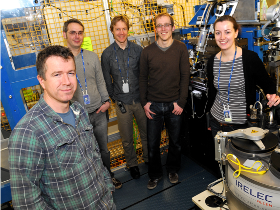
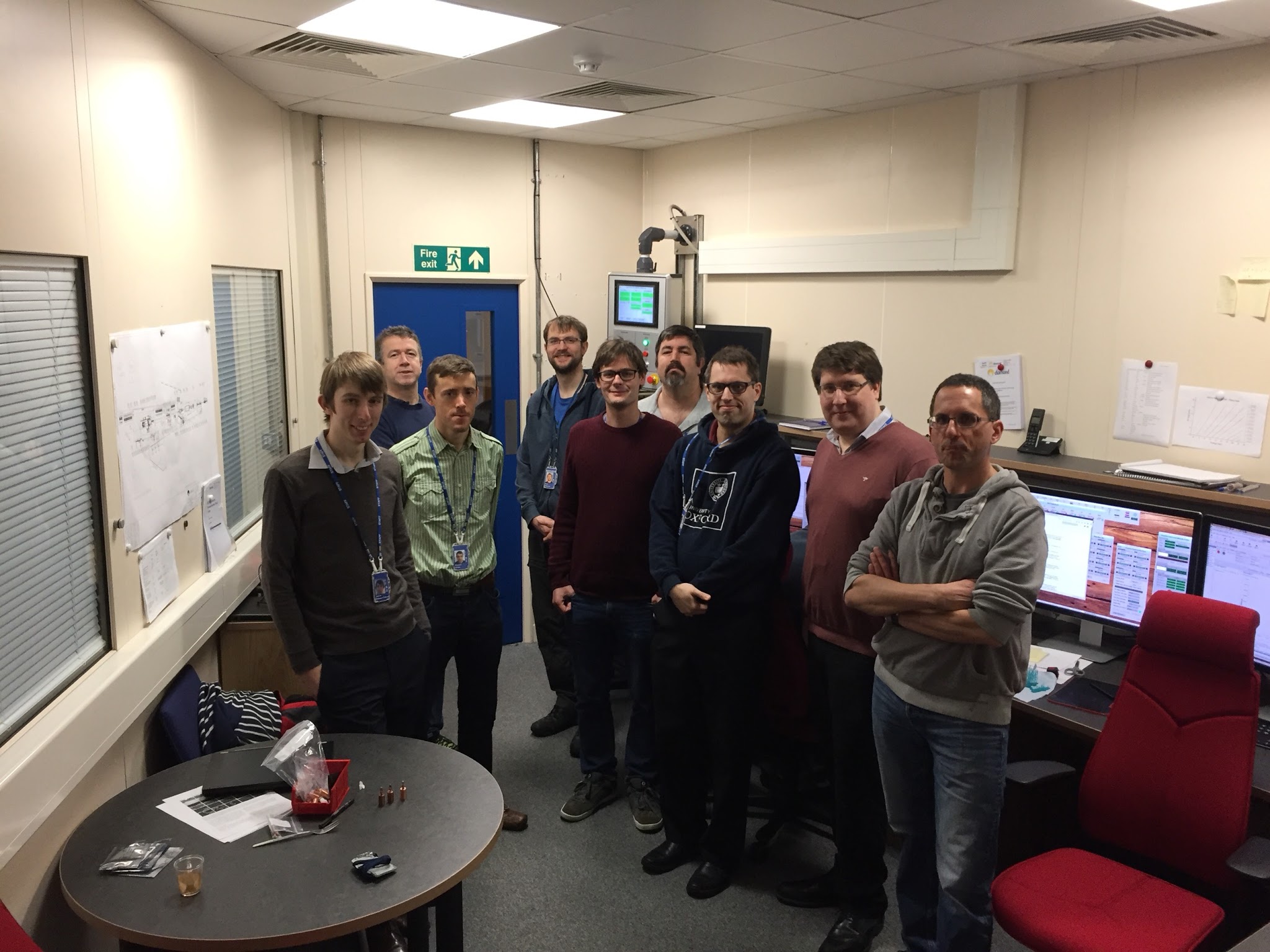
- Figure 3: I24 Team
The work that we did through the MPL benefitted more than just membrane protein crystallography (Axford et al., 2015).In fact virus crystallography had been identified as a clear beneficiary and Dave Stuart’s group became key users and collaborators of the I24 and its team 6,7 (Figure 3). The in situ set up, although demonstrated as being potentially useful for membrane proteins, was in fact wonderfully suited to virus crystallography and the next few years exploited this advantage, up until the cryoEM revolution. Around this period XFEL serial crystallography took off 8 and we at I24 began to understand the limits of synchrotron crystallography as it was in 2010 and clearly recognised the benefits of XFELs for ‘zero-dose’ and ultrafast time resolved measurements. The small crystal limit of synchrotrons was also explored with the potential benefits of XFELs for micron-sized sample becoming evident.
VMXm and DIALS
With the success of I24 and the growing interest in in situ crystallography a case was made by the community for a Phase III beamline facility that could offer additional microbeam capability to address I24 demand, alongside in situ crystallography that could exploit a resurgent interest in room temperature structures. The VMX proposal was put before the Diamond SAC and the project started in 2012 with me leading the microbeam element (VMXm) and Thomas Sorensen taking the lead on the in situ instrument (VMXi).
- Figure 4: First light at VMXm. Graham Duller at the helm looking for light up the beamline.
My experience at I24, my recognition of the practical limits of a beamline like I24 (sample mounted in air using a cryostream to cool the sample), and a good sense of the theoretical possibilities of synchrotrons for MX 9 led to the shaping of the VMXm conceptual design. We were going to leverage the experience of Armin Wagner and his team at I23 who were building a fully in vacuum end-station, and work closely with the Diamond Optics and Metrology group to build a sub-micron beamline for MX that could surpass anything currently in existence when it came to measuring high quality data from a small number of micron-sized crystals. The key words were sample efficiency and data quality. I worked very closely during the design and construction of VMXm with many of Diamond expert support teams. Many of the initial concepts used for VMXm came through discussions with Graham Duller (Figure 4) from the Engineering group. Importantly, we decided very early on in the build of VMXm to include representatives of the machine group and specifically involved the Diagnostics group leader (then Guenther Rehm) to leverage specific expertise on beam stability and control. We created an Integrated Design for Stability Group (IDSG) to steer the design of VMXm throughout the project. This decision was rewarded after we took first light and observed the exquisite beam properties at the sample 10.
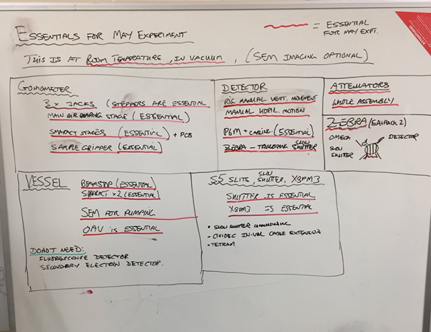
- Figure 5: List of essentials at the beamline to conduct our first ever diffraction experiment at VMXm on a graphene wrapped lysozyme crystal.
Our first experiment was conducted at room temperature with a crystal wrapped in graphene11 and was cobbled together in many respects but gave us confidence we were on the right path (Figure 5). On one of our more challenging test samples (CPV17) where crystals were only 1.2 µm across, we were able to record a complete data set and solve the structure by molecular replacement combining fewer than 20 crystals to the same resolution as the structure determination from XFEL data (LCLS) where around 6000 crystal hits had been used 12 (many more crystals had actually been fired across the sample interaction point but missed the beam!).
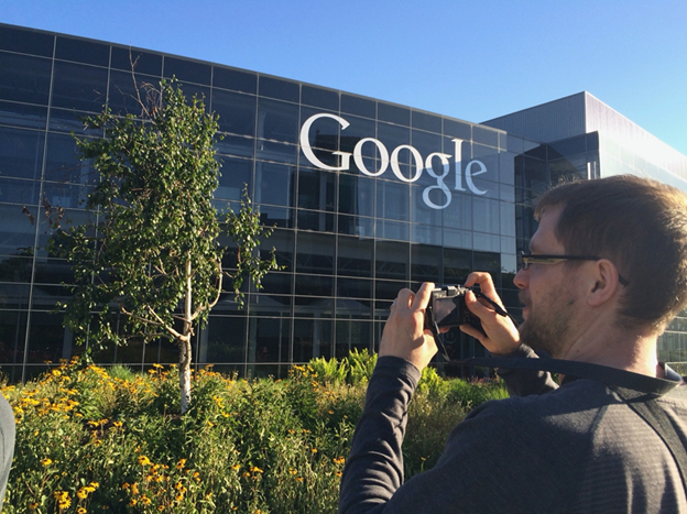
- Figure 6: David Waterman goggling Google during one of our exchange trips with the Berkeley team.
Back in 2012 a thought struck me: The future of data analysis software available to the community at the time was at risk and the lead time for making new developments or analysis methods available to users was prohibitively slow. This was evident every time a new detector was implemented at one of our beamlines that required software updates or there was some identified need for new analysis features. The software suites that I was most familiar with at the time, MOSFLM/SCALA (from CCP4), XDS, D*TREK and HKL (denzo), were either commercial and closed source; open source but with an aging code base and limited development possibilities; or the domain and life’s-works of single developers with limited resources to make substantial changes or improvements. My strong feeling at the time was that there was a substantial gap in the market for a data analysis package that was open source, collaborative and not dependent on a single developer, and could serve as a vehicle to train up a new generation of methods experts to build upon the work of the earlier generations. This idea had been cemented by the work of my then post-doc David Waterman (Figure 6) who had been working on improving the error estimates from integrated intensities and making modifications to MOSFLM to implement the ideas 13.
- Figure 7: DIALS east developers
It was clear then that in order to develop novel methods in a straightforward way it would be essential to have a set of development tools focused around data analysis. I found allies to my thinking at Diamond (Graeme Winter and Dave Stuart), at CCP4 and Cambridge (David Waterman, Andrew Leslie and Garib Murshudov) and across the ocean in Berkeley, CA (Nick Sauter) and in 2012 I made a pitch to CCP4 for the development of a new software package, DIALS. We held the first of many DIALS workshops in June 2012 bringing together many of the key developers in the field (Figure 7). (The development of DIALS was initially funded by the European Union FP7 BioStruct project and later by Wellcome Trust from 2016 to 2025. Their support along with the unwavering support of Diamond, CCP4 and of our collaborators in Berkeley allowed DIALS to flourish over the next decade. I feel very proud when looking at the new generation of developers (Figure 4) who have been trained by developing in DIALS and I feel strongly that the initial ambition of DIALS back in 2012 has been exceeded 14–20.
HeXI and final thoughts
My last major project involvement at Diamond was with HeXI. The concept for HeXI had really been born out of what we had achieved with VMXm, at a time where there was a growing interest in the field for electron diffraction. However, the revitalization of electron diffraction was a classic example of two worlds colliding: that of electron microscopy and X-ray crystallography. My personal view (I was not alone) was that the measurement of high-quality electron diffraction data was not going to be realized by using traditional electron microscopes designed primarily for imaging. Moreover, discussions with Jan Pieter Abrahams (University of Basel) had inspired me to consider the benefits of using high energy electrons to increase sample penetration and potentially improve data quality further for smaller samples. Wellcome Trust again put their confidence in Diamond and in collaboration with the Rosalind Franklin Institute and the MRC-LMB in Cambridge we were awarded funding for HeXI through the Electrifying Life Sciences strategic grant from Wellcome. Alistair Siebert was appointed PBS for HeXI and is now driving the project.
During the development of I24, the MPL and BLEND, VMXm, HeXI and DIALS I have managed to assemble teams of remarkable people at Diamond to work with me. I have also been supported by Diamond users and my brilliant colleagues at other synchrotrons. The world of synchrotron macromolecular crystallography is a very small community. There is naturally some inter and intra-facility competition, but my personal experience is that most people are completely willing to share ideas, give feedback, criticize constructively, learn from each other and be a part of the broader evolution of technology that only happens when everyone pushes in the same direction.
Long may it continue.
Acknowledgements
There are too many people to thank individually. I must however recognize the many beamline builders and software developers who I have learned from over the years. I have been very fortunate to have rubbed shoulders with some incredibly talented and knowledgeable people. But most importantly, if you have worked on or supported any of the projects I discussed here, even in the smallest way, then I owe you a huge thank you. It has been a massive team effort, and I think we have achieved some amazing things together.
References
1. Hendrickson, W. A., Horton, J. R. & LeMaster, D. M. Selenomethionyl proteins produced for analysis by Multiwavelength Anomalous Diffraction (MAD): a vehicle for direct determination of three-dimensional structure. EMBO Journal. 9, 1665–1672 (1990).
2. Moraes, I., Evans, G., Sanchez-Weatherby, J., Newstead, S. & Stewart, P. D. S. Membrane protein structure determination — The next generation. Biochimica et Biophysica Acta (BBA) - Biomembranes 1838, 78–87 (2014).
3. Foadi, J. et al. Clustering procedures for the optimal selection of data sets from multiple crystals in macromolecular crystallography. Acta Crystallographica Section D 69, 1617–1632 (2013).
4. Aller, P., Geng, T., Evans, G. & Foadi, J. Applications of the BLEND Software to Crystallographic Data from Membrane Proteins. Advances in Experimental Medicine and Biology vol. 922 (2016).
5. Axford, D. et al. Structure determination of an integral membrane protein at room temperature from crystals in situ. Acta Crystallogr. D Biol. Crystallogr. 71, (2015).
6. Wang, X. et al. A sensor-adaptor mechanism for enterovirus uncoating from structures of EV71. Nat. Struct. Mol. Biol. 19, (2012).
7. Axford, D. et al. In situ macromolecular crystallography using microbeams. Acta Crystallogr. D Biol. Crystallogr. 68, (2012).
8. Chapman, H. N. et al. Femtosecond X-ray protein nanocrystallography. Nature 470, 73–7 (2011).
9. Holton, J. M. & Frankel, K. A. The minimum crystal size needed for a complete diffraction data set. Acta D66, 393–408 (2010).
10. Warren, A. J. et al. VMXm – A sub-micron focus macromolecular crystallography beamline at Diamond Light Source. J. Synchrotron Radiat. 31, (2024).
11. Warren, A. J. et al. In vacuo X-ray data collection from graphene-wrapped protein crystals. Acta Crystallographica Section D 71, 2079–2088 (2015).
12. Ginn, H. M. et al. Structure of CPV17 polyhedrin determined by the improved analysis of serial femtosecond crystallographic data. Nat Commun 6, (2015).
13. Waterman, D. & Evans, G. Estimation of errors in diffraction data measured by CCD area detectors. J. Appl. Crystallogr. 43, 1356–1371 (2010).
14. Waterman, D. G. et al. Diffraction-geometry refinement in the DIALS framework. Acta Crystallogr. D Struct. Biol. 72, (2016).
15. Beilsten-Edmands, J. et al. Scaling diffraction data in the DIALS software package: algorithms and new approaches for multi-crystal scaling. Acta Crystallogr. D Struct. Biol. 76, (2020).
16. Clabbers, M. T. B., Gruene, T., Parkhurst, J. M., Abrahams, J. P. & Waterman, D. G. Electron diffraction data processing with DIALS. Acta Crystallographica Section D 74, 506–518 (2018).
17. Parkhurst, J. M. et al. Robust background modelling in DIALS. J. Appl. Crystallogr. 49, (2016).
18. Waterman, D. G. et al. Diffraction-geometry refinement in the DIALS framework. Acta Crystallographica Section D 72, 558–575 (2016).
19. Brewster, A. S. et al. Improving signal strength in serial crystallography with DIALS geometry refinement. Acta Crystallogr. D Struct. Biol. 74, (2018).
20. Parkhurst, J. M. et al. Dxtbx: The diffraction experiment toolbox. J. Appl. Crystallogr. 47, (2014).

Diamond Light Source is the UK's national synchrotron science facility, located at the Harwell Science and Innovation Campus in Oxfordshire.
Diamond Light Source Ltd
Diamond House
Harwell Science & Innovation Campus
Didcot
Oxfordshire
OX11 0DE
Copyright © Diamond Light Source. Diamond Light Source® and the Diamond logo are registered trademarks of Diamond Light Source Ltd
Registered in England and Wales at Diamond House, Harwell Science and Innovation Campus, Didcot, Oxfordshire, OX11 0DE, United Kingdom. Company number: 4375679. VAT number: 287 461 957. Economic Operators Registration and Identification (EORI) number: GB287461957003.