

 Industrial Liaison Office
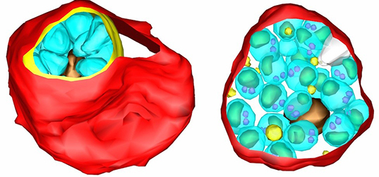
Cryo-soft X-ray Tomography (cryo-SXT)
This relatively new technique provides unique capabilities to capture high resolution (25nm) images of cellular architecture. By maintaining the native state of the samples, this method enables you to investigate the intricate molecular interactions within whole cells to observe biological activities such as viral replication, bacteria clearance, parasite egress, sporulation and many more in unprecendented detail.
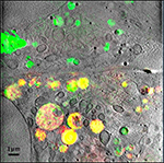
This combination of X-ray tomography with high resolution fluorescence microscopy (co-located with the X-ray instrument) provides a correlative microscopy approach to identify and locate biological structures at the intra-cellular level.
Benefits
- Enables users to study the ultrastructure of vitrified whole cells
- Provides the capability to capture cellular images to a resolution of 25-40 nanometres.
- Allows investigations of molecular interactions in cellular environments
- Enables users to study cells at near-native states (no fixation, staining or micro-sectioning required)
- Facilitates the study of relatively thick biological samples
- Allows longer exposures and serial imaging without significant radiation-induced sample deterioration.
- Integrates with complementary techniques such as cryo-EM and light microscopy modalities.
Near Native Cell Imaging
- Generates 3D slices through the sample
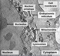
- Collects data on intact and unstained cells through entire cell thickness (up to 8 μm)
- Probes a wide range of sample types from bacteria to whole mammalian cells
Structural Cell Biology
- Studies the effect of drugs on living cells
- Follows the effects of biological processes on the cell e.g. starvation
- Monitors cell regulation processes
- Investigates impact of nanoparticles on cells
Cell Infection Process
- Studies cell infection
- Images viral particles within infected cells
- Examines changes to cell organelles post infection
- Explores host-pathogen interactions
Functional Studies of Cellular Processes
- Performs correlative studies with cryofluorescence microscopy
- Reveals localisation of fluorescent proteins within complex organelles
- Captures processes of dynamic membrane trafficking.
