Better diagnosis for brain cancer
Feb 4, 2021
Feb 4, 2021
Gliomas constitute the vast majority of primary brain tumours and unfortunately, patients diagnosed with gliomas have poor clinical prognoses. Recently, a UK wide team of researchers used Diamond to study the protein IDH1, which is present in all gliomas. The researchers developed a method based on infrared radiation (IR) to determine if the IDH1 had a specific mutation: a mutated version of the IDH1 gene - if present in a glioma - is associated with a better clinical outcome. The current way to determine whether a glioma has the mutation now is to take a biopsy, which is an incredibly dangerous and invasive. A further complication is that several different biopsies may have to be taken across the entire tumour as the mutations are often not evenly distributed. Standard procedure is for the biopsy to be processed and stained in a lab for visible analysis, which means it takes longer to make a diagnosis.
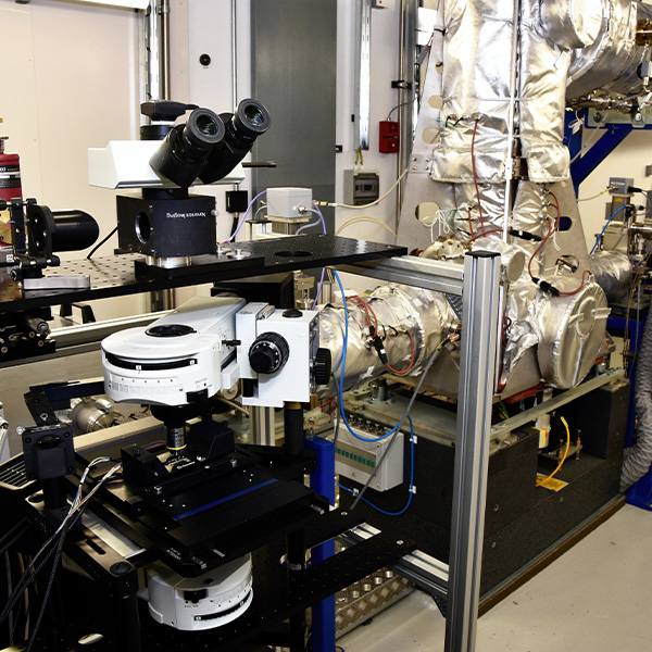
The research team used Diamond’s beamline B22: Multimode InfraRed Imaging And Microspectroscopy (MIRIAM) and other methods broadly classed as “vibrational spectroscopy”. In particular, Fourier-transform infrared spectroscopy (FTIR) rely on the fact that molecules vibrate uniquely when excited by the infrared light, a sort of infrared fingerprint. This means that by measuring the infrared absorption spectra from a tissue, the molecular components (e.g. type of proteins, lipids or small compounds) can be readily identified.
The research team studied by infrared the wild-type protein IDH1 and compared it to the mutant. They discovered that the mutant and the wild-type proteins indeed had different infrared spectral signatures, which could be reliably differentiated. They confirmed these results on biopsy sections from 79 glioma patients with both sensitivity and specificity significantly above 80%.
This finding means that in the future, it might be possible for surgeons to use FTIR analysis to get a real-time read out of the kinds of mutations present in the gliomas of their patient. This could then help them make timely medical decisions to deliver personalised medicine, i.e. treatment specific to the patient to give them the best possible outcomes.
The research study also investigated whether blood serum could be analysed to give clinical information on gliomas. They wanted to know if they could measure any changes in the blood of patients that was linked to the mutation in the IDH1 enzyme. For this, blood serum samples from an additional 72 patients were collected. The research team discovered a method for processing the blood sample and accurately determining which variant of the IDH1 protein a patient had 69% of the time.
This exciting scientific finding provides clinicians with a relatively non-invasive way to better understand the brain tumours of individual patients. This gives them important information to administer more personalised treatments and decide on the kind of surgical intervention that might be needed.
While the study used a synchrotron as a radiation source, it doesn’t mean that this is the only way to do FTIR, which can also be carried out via benchtop systems. Synchrotron IR has a number of advantages, like being up to 1,000 times brighter than conventional benchtop source, while still being non-damaging to organic matter, and a vastly wider broadband than a typical laser source, for example. In practice, synchrotron IR is ideal for spectroscopy in exploratory research like this and allows high quality data even at the spatial scale of a cell in tissues. This combination of IR spectroscopy and microscopy allows researchers to look at smaller sections of human tissues with superior spectral sensitivity necessary for global analysis and refinement of the method.
Gianfelice Cinque, Principal Beamline Scientist at Diamond, said:
Synchrotron based FTIR has proven to be key in proof-of-concept biomedical studies. On this basis, it becomes easier to adapt IR microspectroscopy methods to lab instruments and focus only on the molecular differences that are important and were validated by synchrotron FTIR at Diamond, for example.
Matthew Baker, from University of Strathclyde and ClinSpec Diagnostics Ltd, said:
Our research has further demonstrated the possible patient benefit that can be achieved from clinical spectroscopy. We are now working to translate these research advances into the clinic so we can positively affect the lives of patients worldwide.
To find out more about B22: Multimode InfraRed Imaging And Microspectroscopy (MIRIAM) at Diamond, or to discuss potential applications, please contact B22 Principal Beamline Scientist, Gianfelice Cinque: [email protected]
James M. Cameron et al. Interrogation of IDH1Status in Gliomas by Fourier Transform Infrared Spectroscopy Cancers. DOI: 10.3390/cancers12123682 12 3682 (December 2020)

Diamond Light Source is the UK's national synchrotron science facility, located at the Harwell Science and Innovation Campus in Oxfordshire.
Diamond Light Source Ltd
Diamond House
Harwell Science & Innovation Campus
Didcot
Oxfordshire
OX11 0DE
Copyright © Diamond Light Source. Diamond Light Source® and the Diamond logo are registered trademarks of Diamond Light Source Ltd
Registered in England and Wales at Diamond House, Harwell Science and Innovation Campus, Didcot, Oxfordshire, OX11 0DE, United Kingdom. Company number: 4375679. VAT number: 287 461 957. Economic Operators Registration and Identification (EORI) number: GB287461957003.