Thin multilayer structures comprising of thin layers of alternating elements or compounds find widespread technological applications – be it the anti-reflection coating in the visible range or the waveguide structures for X-rays. In the X-ray regime they are also used in many technological applications such as X-ray astronomy, microscopy, spectroscopy, and as filters and monochromators for synchrotron radiation and free electron X-ray lasers. It is important to correlate the measured optical properties of such multilayer systems with their structures. It is however not a trivial matter to perform the full structural characterisation of such thin multilayer structures. We show that the structural characterisation of both the high and low z (atomic number) material layers of thin periodic multilayer structures can be performed independently by measuring the X-ray standing wave enhanced elastic and inelastic scattering intensities. The method has been tested by analyzing an Nb/C/Nb trilayer and a Mo/Si periodic multilayer structure. We demonstrate that the present approach is especially suitable for studying multilayer structures comprising low atomic number layers, because it eliminates the requirement of a fluorescence signal, which is very weak in the case of low z materials.
Thin layered structures1, 2 find a wide range of applications in many fields, as they offer unique structural, magnetic and electronic properties. The micro-structural properties such as layer thickness, interface roughness, interlayer formation etc. of such layered materials is usually determined from X-ray reflectivity (XRR), X-ray diffuse scattering or X-ray standing wave (XSW) induced fluorescence measurements. The more commonly used XRR technique however has the limitation that it does not provide any element-specific information. Moreover, it only gives the composite thickness and roughness values for the high and low z layers of any periodic multilayer structure, and sometimes it becomes rather difficult to establish the true micro-structural parameters of such structures. XSW induced fluorescence measurement is a more direct method to determine the structural properties of high z layers in periodic multilayer structures. The fluorescence signal from the low z layer however is weak due to its low fluorescence yield and the strong self absorption in the layered material. Therefore, the structural information of the low z layers is determined indirectly from the XSW fluorescence measurement of the high z layers.
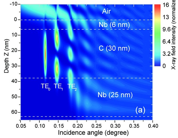

Figure 1: (a) Contour plot of x-ray field intensity distribution inside an Nb (6 nm)/ C (30 nm)/ Nb (25 nm) trilayer structure as a function of incidence angle and the depth Z in the layer medium, at X-ray energy of 17 keV. (b) Variation of the elastic, Compton and photoelectric X-ray cross sections of Nb and C as a function of incident X-ray energy.
The scattered X-rays (elastic and Compton) emitted from a thin layered material also contain the information about structural parameters of the thin film system. In practice, however, it is very difficult to retrieve such information. In literature, no direct formulation has been provided to determine the structural properties of the layered materials using XSW enhanced elastic and Compton scattered X-rays. In some applications, it is important to determine the layer properties of the individual layers, for example, in X-ray wave guide structures (trilayers) where a thick low z layer is sandwiched between two high z layers. It is well know that the Compton and elastic X-ray scattering cross sections largely depend on the atomic number (z) of a material (for example: photoelectric absorption a z4, elastic scattering a z2, and Compton scattering µaz). We have used this property to correlate the structural properties of high and low z material layers with their measured angle-dependent XSW enhanced elastic and Compton scattering profiles. Unlike conventional XSW fluorescence measurements, the method proposed by us has the advantage that one can determine the information on both high and low z layers independently.
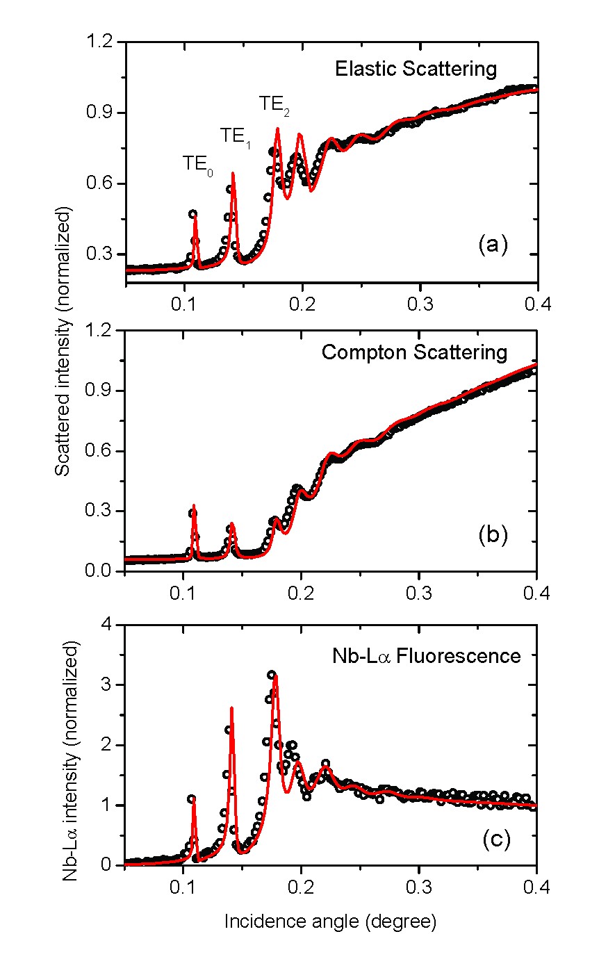
Figure 2: Measured and fitted elastic, Compton, and Nb La fluorescence profiles from an Nb/C/Nb trilayer structure. The scattered points show the experimental data while solid lines show the best fits to the measured profiles.
To demonstrate the capability of the method, two repetitive multilayer structures – an Nb/C/Nb trilayer structure and a Mo/Si periodic multilayer structure have been analysed. The results of the former system are presented here. Measurements were performed on the B16 Test beamline. In the Nb/C/Nb trilayer structure, a low z layer (carbon) is sandwiched between two high z (niobium) layers. Figure 1(a) gives the contour plot of X-ray field intensity distribution inside the Nb (6 nm)/C (30 nm)/Nb (25 nm) trilayer structure, calculated at an incident X-ray energy of 17 keV, as a function of incidence angle and depth Z in the layer medium. From Figure 1(a) it can be seen that various transverse electric field modes (TE0, TE1, TE2, etc) are excited in the C layer medium at definite incidence angles. At large incidence angles (for higher modes), the X-ray field intensity moves towards the interfaces of the layered medium, thus providing the possibility to determine the interface properties of the high and low z layers by measuring the fluorescence or scattered intensities from the layered medium. In Figure 1(b) we have shown the variation of various X-ray cross sections as a function of X-ray energy, for Nb and C elements. It can be seen that different cross sections have strong photon energy dependence, and also a z-dependence. The figure shows that one can get enough contrast of different X-ray cross sections between high z (niobium) and low z (carbon) materials.
Figure 2 shows the measured and fitted elastic, Compton and X-ray fluorescence profiles of the Nb/C/Nb structure. From Figures 2(a) and (b) it can be seen that the elastic and Compton scattering profiles are quite different from each other, especially at incidence angles below the critical angle of the Nb layers (?c ~ 0.18°). For instance, the relative peak height ratios of the TE0, TE1 and TE2 modes are quite different, as this ratio strongly depends on the thickness ratios of the high and low z layers. Moreover, at large incidence angles, the higher transverse electric field modes (i.e. TE1, TE2, etc) move towards the top and bottom Nb cladding layers (Fig. 1a). As a result, the peak intensity of these modes increases in the measured elastic scattering profile while it decreases inversely in the measured Compton profile. The Compton scattered intensity for higher electric field modes decreases because the effective amount of XSW intensity contained in the low z medium (carbon layer) decreases. Figure 2(c) shows measured and fitted Nb La fluorescence profiles for the Nb/C/Nb trilayer structure. From Figures 2(a) and (c) it can be seen that the measured elastic scattering and fluorescence profiles show more or less similar behavior. The Compton scattering contribution arises largely from the low z material; hence the Compton scattering profile offers a more accurate determination of structural properties of the C layer. On the other hand, elastic as well as Nb La fluorescence profiles originate from the high z layers and therefore facilitate precise determination of structural parameters of the Nb layers. By analyzing all the three profiles, we have determined the thickness of the top and bottom Nb cladding layers to be 6.5 nm and 25.0 nm, respectively. The thickness of the carbon layer was found to be 30.7 nm. The structural parameters derived from scattering and fluorescence measurements were then used to fit the X-ray reflectivity profile, which was found to match closely with the measured reflectivity data (Fig. 3).
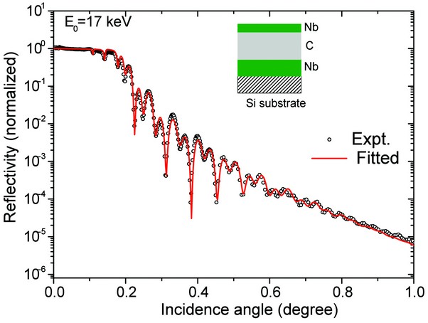
Figure 3: Measured XRR profile of Nb/C/Nb trilayer structure at incident x-ray energy of 17 keV. The solid red line shows the fitted XRR profile using the structural parameters derived from the best fit results of elastic, Compton, and Nb La fluorescence measurements.
The technique presented here offers the advantage that by controlling the incidence angle, one can control the depth of the standing wave field pattern, and hence the probed depth inside any thin film medium. On the other hand, the photon energy of the detected X-ray radiation (elastic and Compton scattered as well as fluorescent X-rays) provides elemental sensitivity. This independent control of spatial and elemental sensitivity makes the technique a powerful material characterisation tool.
In conclusion, we have shown that the Compton and elastically scattered X-rays from thin multilayer structures can be exploited to determine structural properties of both high and low z materials, thereby facilitating complete characterisation for thin layered materials. It greatly overcomes the limitation of the conventional XSW fluorescence technique, where analysis of the low z layers is a challenge, and makes possible the analysis of both high and low z layers independently. The method therefore is especially suitable for the characterization of multilayer structures comprising of low z layers (e.g. Langmuir-Blodgett films, thin polymer or biological enzyme sensor films).
Tiwari, M.K. and Sawhney, K.J.S. Structural characterization of thin layered materials using X-ray standing wave enhanced elastic and inelastic scattering measurements. J. Phys.: Condens. Matter. 22, 175003 (2010)
References
- Spiller, E. Appl. Phys. Lett. 20, 365 (1972).
- Barbee, T.W. Synthetic modulated structure materials. Academic Press, New York. pp. 313 (1985).
