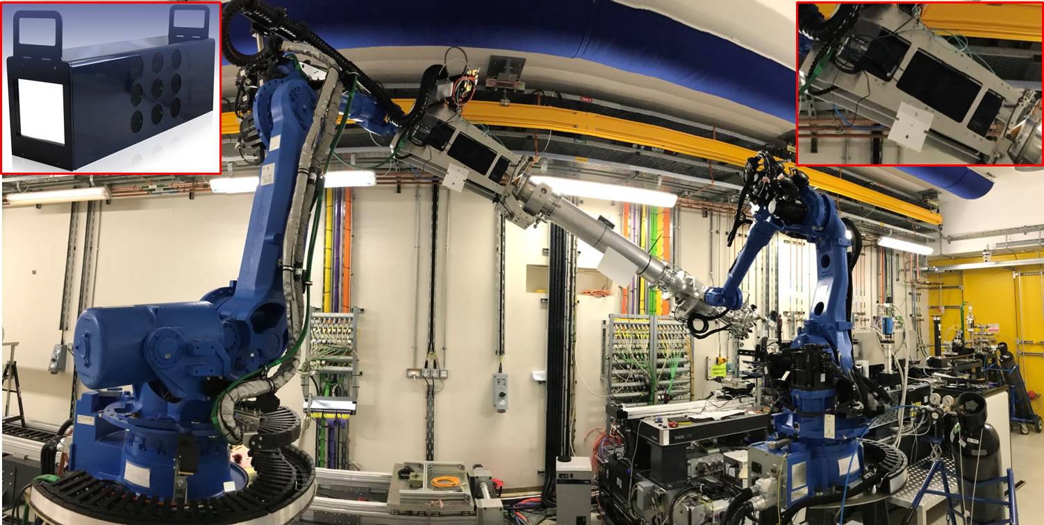
Image: The ExcaliburRX-3M detector mounted on the robot arm on beamline I13. Inset right, zoomed portion of the image showing the detector in the mounting cage. Inset left, a bench-top photograph of the ExcaliburRX-3M.
The ExcaliburRX detector was designed specifically for the requirements of the I13 Coherence beamline for experiments using techniques such as ptychography, ptychotomography and coherent X-ray diffraction. For ptychography and ptychotomography, the detector can be mounted on a 15m long translation rail, enabling sample-to-detector distances of up to 15 metres. For Bragg coherent diffraction imaging, the detector can be mounted on a robot arm allowing to place the detector at a particular Bragg angle. The image above shows the detector mounted on the robot arm assembly. The detector is central and to the top of the image, and is mounted in a custom fabricated cage at the end of a short length of beam pipe.
These unique capabilities necessitated the construction of a large area detector with a small pixel size. The Medipix3RX ASIC with 256x256 55 um pixels and three-side-buttable format was chosen to meet these requirements. Information about the available form factors can be found in Detector Details, and more information about the key technologies used are available in the Sensor Module and Front End Module pages.
A number of commissioning experiments were performed on the I13 beamline, in order to both calibrate the detector and to show the capabilities for some readily available, but representative, use-cases.
Direct Absorption Imaging of a Computer Mouse Peripheral
The first experiment performed was direct absorption imaging of a computer mouse peripheral using a microfocus beam. A pressed powder pellet of potassium bromide was placed behind an aperture and at the focal point of a pair of KB mirrors. The 15 keV beam was focussed to a spot size of about 20 um and provided fluorescent X-rays irradiating the computer mouse.
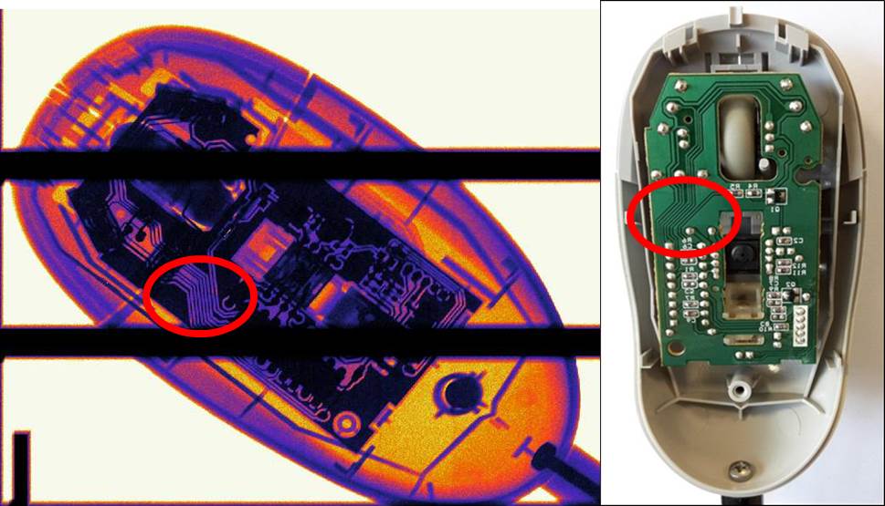
As can be seen from the image, small features such as solder pads and their attached surface-mount electronic components can be clearly seen, along with PCB tracks (highlighted in red). This shows a good resolution of features on the scale of approximately 0.5 mm.
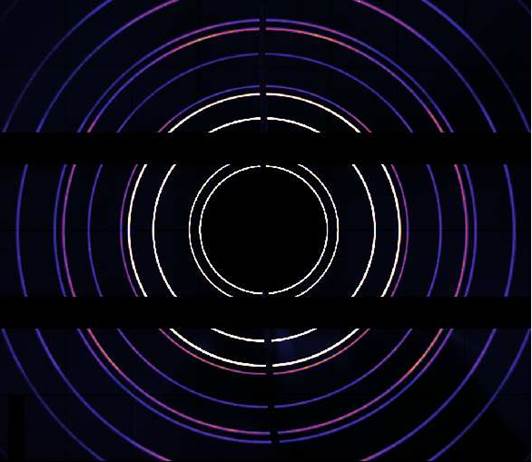
Cerium Oxide Powder Diffraction
- Image from CeO2 powder diffraction
A sample of CeO2 powder was prepared and placed at a distance of around 4cm from the window of the ExcaliburRX-3M detector. Diffraction X-rays then illuminated the detector from a two-theta scattering angle of up to 50 degrees. The sample was distributed evenly about the surface area of a kapton disc, with half masked off during the sample preparation stage. This gave a powder-free region to measure background X-rays from small angle scattering processes which featured quite highly due to the detector being on-axis to the beam. A beam stop wire was centred to absorb any direct beam.
The image to the right shows data taken with a 13 keV beam, following post-processing for background removal.
Siemens Star Ptychography
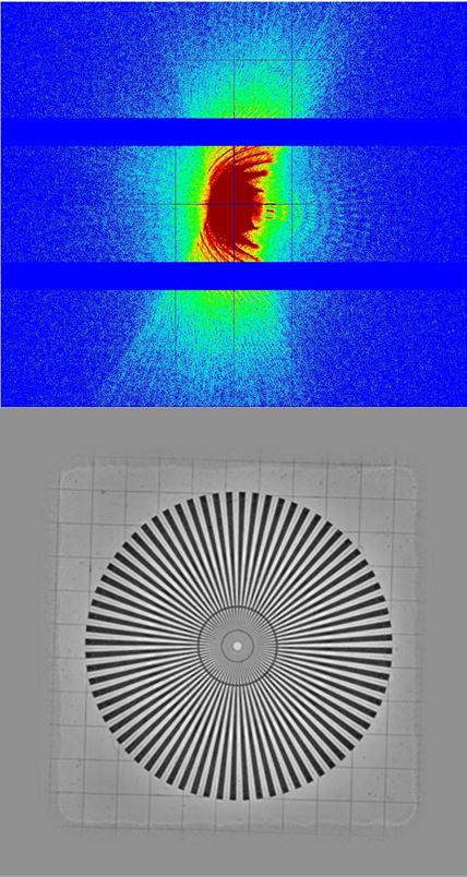
- Sample diffraction frame and reconstructed object phase image of a Siemens Star sample
The ptychography of a Siemens Star sample is a frequently used tool in the process of setting up a beamline for a ptychography and ptychotomography experiment, and provides an excellent readily available but real-world example of a ptychography experiment.
In this commissioning experiment, Frenel-Zone plates were used to focus 9.7 keV X-rays to a spot size of around 25um on the sample. Using step sizes of 1/5 of the size of the beam spot, the sample was rasterized in order to record a series of diffracted images with the ExcaliburRX-3M detector. These images were reconstructed using a pychographic iterative engine algorithm, allowing reconstruction in the object space. In order to obtain an image in the maximum field of view, the detector was placed at 14 m from the sample.
The top panel of the image to the right shows a single frame from this experiment. This shows a single diffraction image, many of which are analysed in the full reconstruction. The lower panel shows the full Siemens Star sample reconstructed from the diffraction data in the object phase, with a field of view of around 200 um obtained. With optimisations, resolutions on the scale of a few 10s of nanometres are achievable with the ExcaliburRX-3M detector installed on the I13 beamline.

Diamond Light Source is the UK's national synchrotron science facility, located at the Harwell Science and Innovation Campus in Oxfordshire.
Diamond Light Source Ltd
Diamond House
Harwell Science & Innovation Campus
Didcot
Oxfordshire
OX11 0DE
Copyright © Diamond Light Source. Diamond Light Source® and the Diamond logo are registered trademarks of Diamond Light Source Ltd
Registered in England and Wales at Diamond House, Harwell Science and Innovation Campus, Didcot, Oxfordshire, OX11 0DE, United Kingdom. Company number: 4375679. VAT number: 287 461 957. Economic Operators Registration and Identification (EORI) number: GB287461957003.