Instruments by Science Group
B23 Contact
Beamline Phone Number:
+44 (0) 1235 778889
Principal Beamline Scientist:
Giuliano Siligardi
Tel: +44 (0) 1235 778425
E-mail: [email protected]
Science Group Leader
Robert Rambo
Email: [email protected]
Tel: +44 (0)1235 56 7675
B23 Circular Dichroism
Status: Operational
Beamsize: 0.5mm² to 2mm x 4mm
Detector: PMT
Wavelength: 125 - 650 nm
Energy: 2 - 10 eV
Detector: PMT
Wavelength: 125 - 650 nm
Energy: 2 - 10 eV
B23: Circular Dichroism
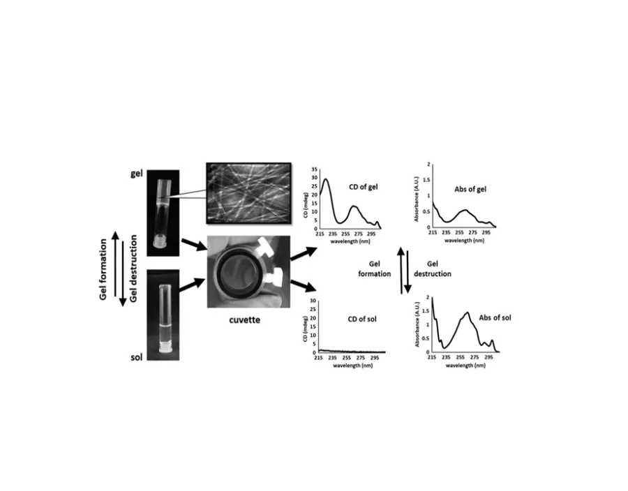
Circular Dichroism (CD) is the spectroscopic technique to study in solution a wide variety of chiral materials such as small molecules (drugs), polymers and biopolymers (nucleic acids, proteins, carbohydrates and lipids). In particular for proteins, knowledge of the structure-function relationship is essential to dissect the mode of action and to identify new targets for novel drug therapeutics.
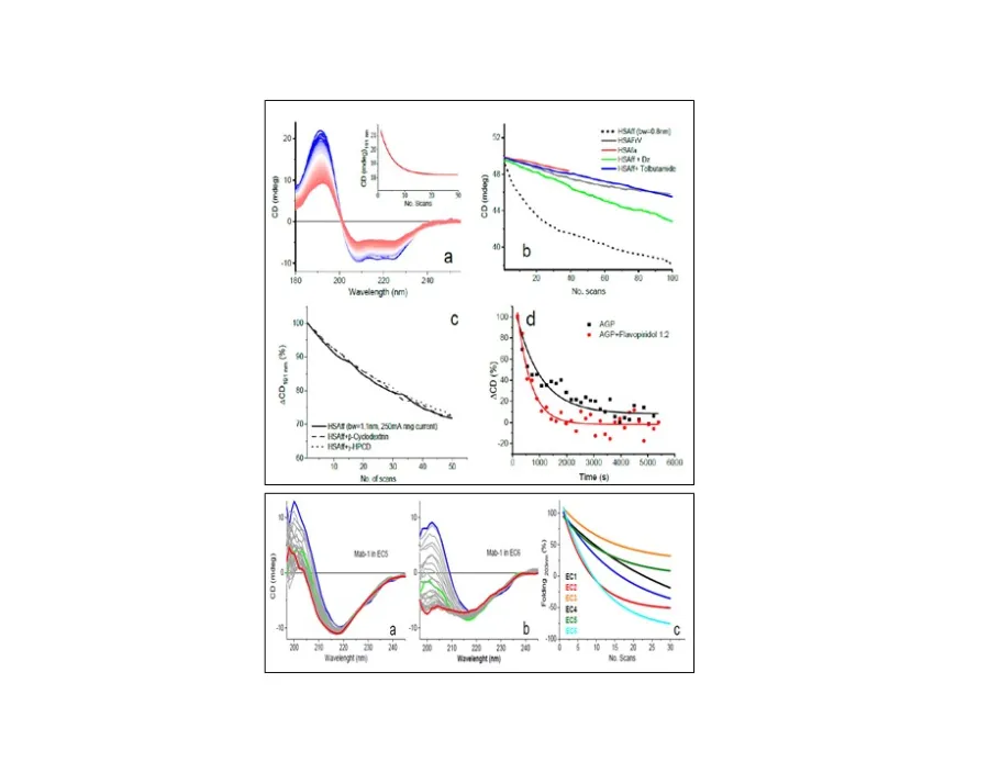
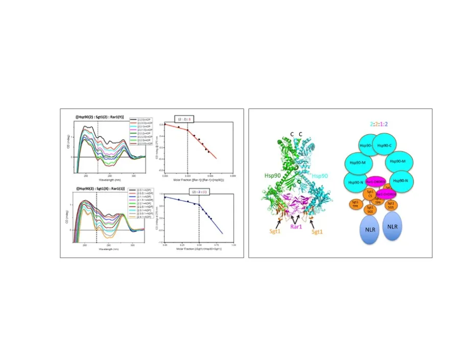
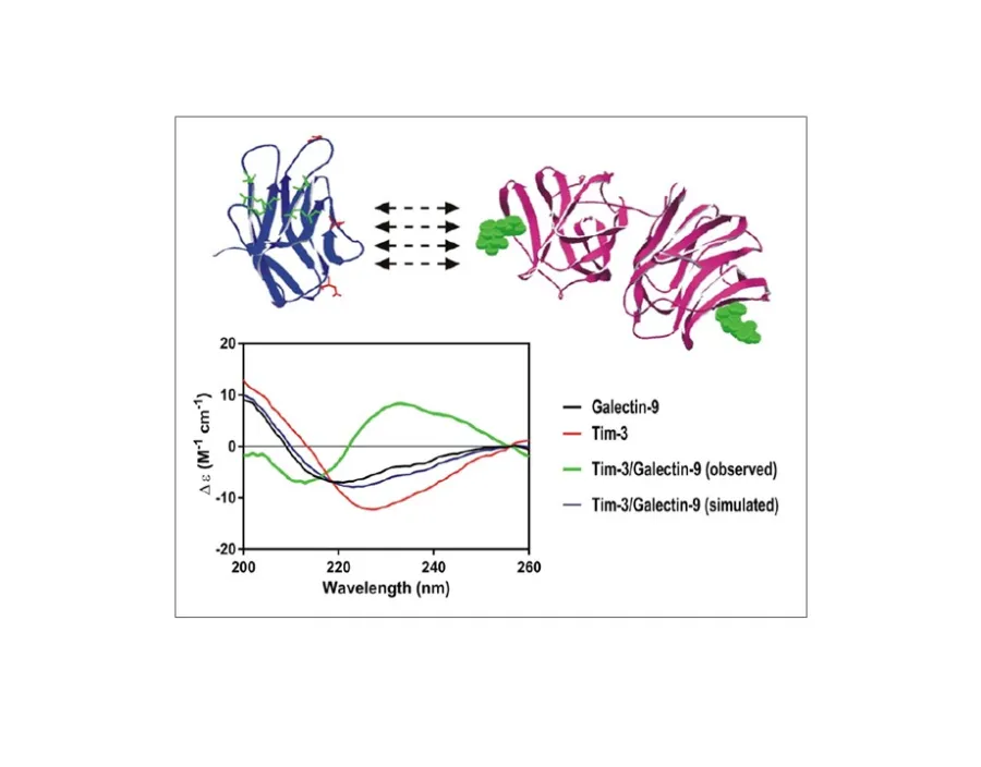
For rigid and well structured systems, like enzymes and globular proteins, CD is a low-resolution technique compared to NMR and X-ray crystallography. However, a third to half of mammalian proteins have natively disordered structures that are unsuitable for NMR and X-ray crystallography. CD is the ideal technique to investigate protein/ligand binding interactions of these important systems involved in signal transduction of normal and tumour cells.
B23 produces a collimated beam of small cross section at the sample (about 1mm (V) x 2mm (H)) enabling the measurement of smaller volumes of sample solutions several times better than commercial CD instruments. High photon flux across the UV region improves the signal-to-noise of CD measurements.
B23 is used by researchers in the biological, biochemical, chemical, pharmaceutical, and crystallographic sciences to examine proteins, nucleic acids, carbohydrates, biopolymers, small ligands and the interactions of these molecules to form macromolecular and drug complexes.
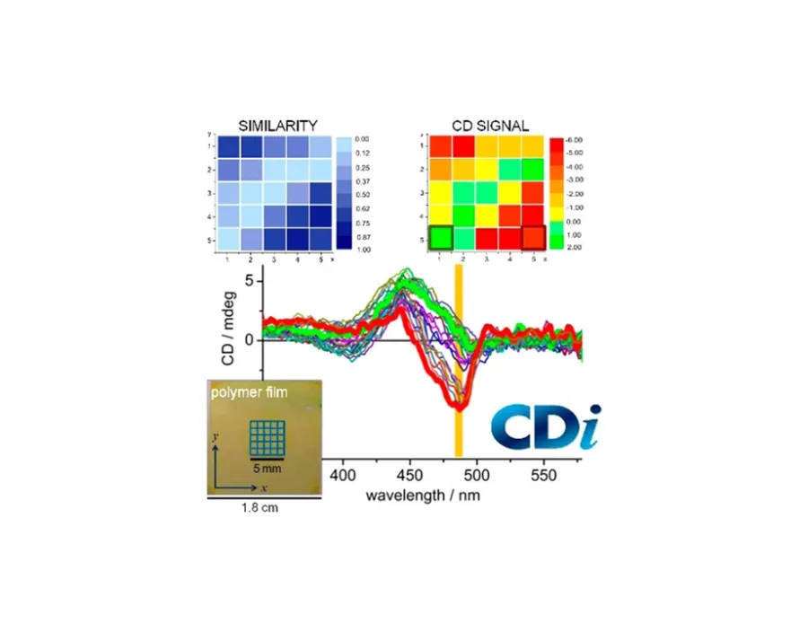
B23 has also the only world state of the art facility, the Mueller Matrix Polarimeter (MMP) on module B end-station to study the homogeneity of the supramolecular structures of thin films of chiral materials deposited on fused quartz substrates. In the solid state, linear anisotropies (linear dichroism and linear birefringence) and photoelastic modulator (PEM) imperfections can distort substantially the CD spectral profile giving misleading interpretations. With B23 MMP the true CD can be extracted at the highest spatial resolution of about 50 micron in mapping mode (usually from 1x1mm to 4x4mm areas) at single wavelength or 190-650nm spectral range. As the chiroptical properties of chiral materials are strictly related to their function and activity, for example optoelectronic materials with photovoltaic or OLED properties, the ability to characterise thin chiral films prepared under a variety of conditions and protocols such as drop cast, spin coating, spray at different temperatures (-170 to +350 C range) and concentrations will enable the determination of the critical parameters essential for obtaining reproducible, uniform and homogeneous specimens.

Diamond Light Source is the UK's national synchrotron science facility, located at the Harwell Science and Innovation Campus in Oxfordshire.
Diamond Light Source Ltd
Diamond House
Harwell Science & Innovation Campus
Didcot
Oxfordshire
OX11 0DE
Copyright © Diamond Light Source. Diamond Light Source® and the Diamond logo are registered trademarks of Diamond Light Source Ltd
Registered in England and Wales at Diamond House, Harwell Science and Innovation Campus, Didcot, Oxfordshire, OX11 0DE, United Kingdom. Company number: 4375679. VAT number: 287 461 957. Economic Operators Registration and Identification (EORI) number: GB287461957003.