____________________________________
Industrial Liaison Group:
Tel: +44 (0) 1235 778797
E-mail: [email protected]
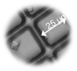
Developments on macromolecular crystallography (MX) beamlines have achieved very high throughput and microfocus X-ray facilities have made it possible to successfully collect diffraction data from samples previously considered too small or too disordered. However, when the use of microfocus beams is coupled with the small size and poor optical properties of some samples, sample alignment and detection becomes a key problem.
Microfocus beamline users generally face one of two challenges prior to successful data collection. The first challenge involves locating small microcrystal(s) within a larger sample mount and alignment of these with the X-ray beam. The other critical issue is the location of well diffracting crystal subvolumes in larger, disordered crystals.
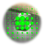
A raster-scanning method has been developed for MX beamlines. A virtual grid is superimposed over all or part of the sample when viewed by an on-axis microscope. A Grid Scan is performed prior to data collection and is done with very low transmission to reduce any effects of radiation damage. Each image is processed and evaluated by a scoring system as shown on the right. All MX beamlines at Diamond are equipped with Grid Scan which is implemented in Diamond’s GDA (Generic Data Acquisition) software.
The Grid Scan is now routinely used on all Diamond’s MX beamlines and is an invaluable tool for the detection and alignment of microcrystals as well as for locating the best diffracting parts of disordered crystals. The application range is further extended for “invisible” crystal samples either in the loops, meshes or plates, where crystals may be embedded within refractive materials of crystal plates, solvents, or within opaque materials as for crystals harvested from the lipid mesophase such as membrane proteins. The technique is also frequently and successfully used for detection of crystals in ice covered loops.


“GPCR crystallography is a highly challenging area of structural biology, where we are dealing with small crystals in an opaque background. The grid scan technique enables us to optimise the beam for each crystal, maximising the rate at which protein crystals can be scanned and the quality of the diffraction data collected.”
Dr Joao Dias, Principal Scientist at Heptares Therapeutics

Diamond Light Source is the UK's national synchrotron science facility, located at the Harwell Science and Innovation Campus in Oxfordshire.
Copyright © 2022 Diamond Light Source
Diamond Light Source Ltd
Diamond House
Harwell Science & Innovation Campus
Didcot
Oxfordshire
OX11 0DE
Diamond Light Source® and the Diamond logo are registered trademarks of Diamond Light Source Ltd
Registered in England and Wales at Diamond House, Harwell Science and Innovation Campus, Didcot, Oxfordshire, OX11 0DE, United Kingdom. Company number: 4375679. VAT number: 287 461 957. Economic Operators Registration and Identification (EORI) number: GB287461957003.

 Industrial Liaison Office