Find out more about our ambitious upgrade project, delivering more brightness, more coherence, and greater speed of analysis to UK science. More about Diamond-II
![]()
Find out more about Diamond's response to virus research.
![]()
A team of scientists from the University of Oxford and the University of Birmingham have just published one of the most comprehensive multi-disciplinary reviews covering nearly 40 years of discoveries and advancements in the study of enamel and its demineralisation (caries). The review reveals how synchrotron radiation facilities - such as Diamond Light Source - enabled unprecedented new insights into dental tissue function and degradation at different scales.

Caries remains a debilitating condition that lacks adequate prevention and treatment that demands further research to find innovative ways to overcome its detrimental impact on global health. The disease had a global prevalence of around 2.3 billion in 2017 (in permanent teeth). In addition to the clinical effects of pain and discomfort, aesthetic issues, and eventually tooth loss, it constitutes a huge economic burden, estimated to be billions of USD worldwide in painful disruptive treatments.
The team’s paper; “Synchrotron X-ray Studies of the Structural and Functional Hierarchies in Mineralised Human Dental Enamel: A State-of-the-Art Review” was published in the Dentistry Journal 10th Anniversary Issue (April 2023). https://doi.org/10.3390/dj11040098. Its strategic aim was to identify and evaluate prospective avenues for analysing dental tissues and developing treatments and prophylaxis for improved dental health.
Team leader, Professor Alexander Korsunsky, Professor and Fellow Emeritus at Trinity College, Oxford, explains;
Understanding the mechanism of caries development requires tracing the pathways of the biological, chemical, and structural processes that unfold progressively from the microbial and crystal level up to the macroscopic scale. This necessarily engenders the need to visualise and understand tissue organisation and function, along with its interaction with the microbial and chemical environment, through static and dynamic studies. Synchrotron-based studies offer unique tools for this purpose, due to the versatile interaction of X-ray photons with the organic and inorganic tissue components.
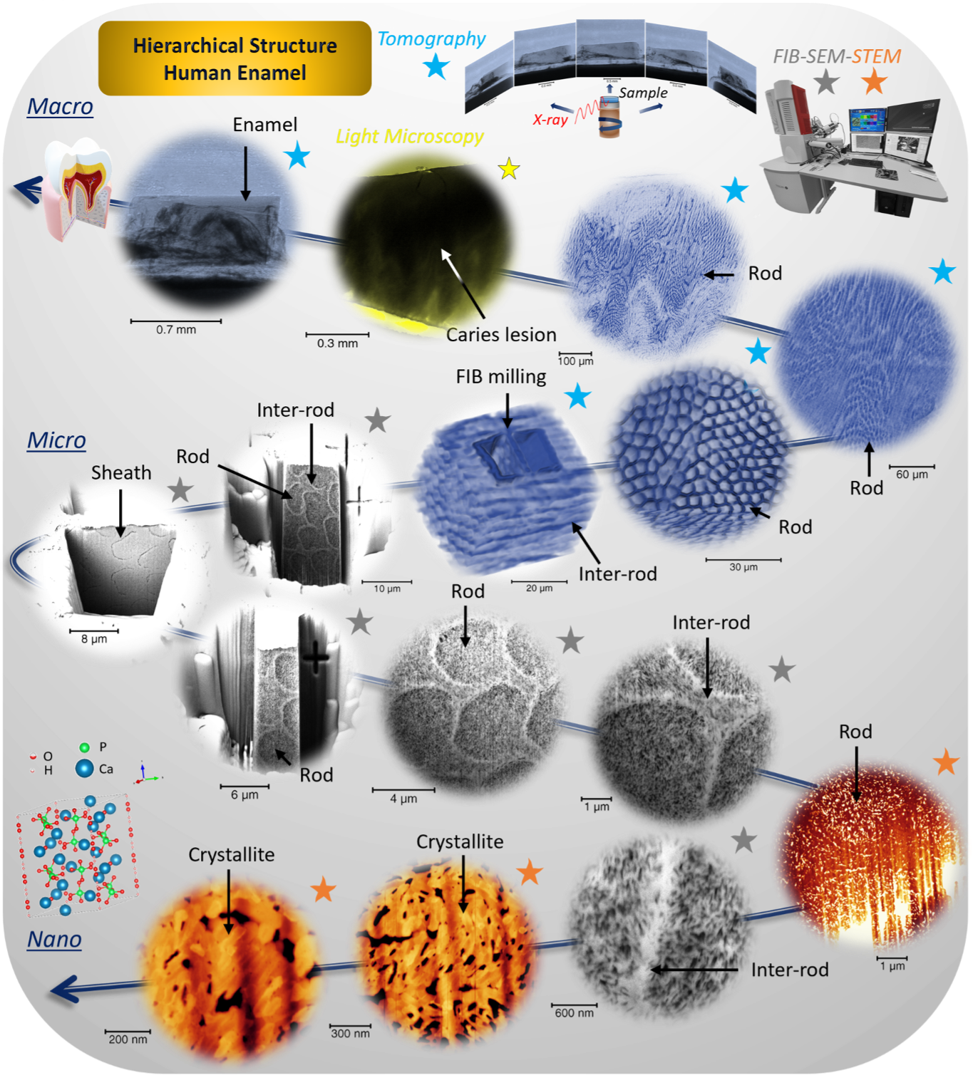
Hard dental tissues possess a complex hierarchical structure that is particularly evident in enamel, the most mineralised substance in the human body. Its complex and interlinked organisation at the Ångstrom (crystal lattice), nano-, micro-, and macro-scales is the result of evolutionary optimisation for mechanical and functional performance: hardness and stiffness, fracture toughness, thermal and chemical resistance. Understanding the physical–chemical–structural relationships at each scale requires the application of appropriately sensitive and resolving probes.
Dr Cyril Besnard, the lead author, adds;
Currently, about 50 synchrotron facilities worldwide are contributing an outstanding amount of research work along with the continuous improvement of analytical approaches. This is due to the fact that synchrotron X-ray techniques offer the possibility to progress significantly beyond the capabilities of conventional laboratory instruments, i.e., X-ray diffractometers, and electron and atomic force microscopes. The last few decades have witnessed the accumulation of results obtained from X-ray scattering (diffraction), spectroscopy (including polarisation analysis), and imaging (including ptychography and tomography).
The first section of the review briefly covers the structure of the enamel (and dentine), describes dental caries disease and its causative factors, including the nature and organisation of biofilm and its effects on the enamel, and discusses the existing strategies for remineralisation. The second section provides an overview of synchrotron facilities, followed by a description of the application of synchrotron methods to dental tissue studies: diffraction (scattering), imaging (including tomography and ptychography), and spectroscopy.
Dr Igor Dolbnya, senior beamline scientist on the B16 Test beamline at Diamond, comments;
The modern synchrotron, like the UK’s Diamond Light Source, offers the versatility of utilizing customised experimental setups, which can be categorised based on the type of detector and relevant setup; the energy in use, either soft or hard X-rays (in vacuum or air or liquid); the presence of magnetic fields or temperature control; and the type of monitoring process (static or dynamic analysis) and equipment. The continuous development of synchrotron facilities, techniques, and devices, means that the future will be bright for the research into mineralised tissues.
The review summarises studies using synchrotron techniques for structural, imaging, and chemical analyses. The utility of these methods is emphasised in terms of bringing new insights, and the benefits of the combined use of multiscale correlative techniques. Diamond’s facilities and beamlines have been used extensively by the authors to study dental tissue and are covered in the review. It is a great example of a multi-disciplinary approach on one research topic as the team used beamlines spread over four different science groups at Diamond, including I08-1, I12, I13-2, I13-1, I14, I18, I22, DIAD, ePSIC, and B16.
Many recent studies are summarised in the review with details and knowledge from state-of-the-art analysis, which could be implemented in future studies. For example, to elucidate the phenomenon of caries and explore avenues such as the 3D structure of the nanocrystallites, the motion of atoms occurring during demineralisation, the in situ process of demineralisation by acid from the bacteria using multimodal imaging, in the time, space and energy domains. The researchers state that these techniques can be applied to design and implement new studies for enamel remineralisation and to develop novel biomimetic materials and strategies to repair enamel and dentine. This direction of research lies at the core of the recently awarded £2.3M EPSRC grant to Prof. Alexander Korsunsky and collaborators entitled “Elucidating the pathways for human tooth enamel mineralisation by 4D microscopy and microfluidics”.
Dr Adrian Mancuso, Diamond’s director of Physical Sciences, concludes;
Synchrotron-based analyses have led to major advances in the structure, and hence mechanical properties, of dental tissues. This includes the caries process, and other dental fields, aiming to improve quality of life. However, there are still open research questions that warrant further investigation. Continuously building on current research will help us to better understand the changes in diseased tissue structure and, in turn, its management.
The review also highlights the importance of the applications and approaches carried out on the research of other materials. The knowledge gathered from these approaches can often be transferred to dental caries research, to bring new research opportunities and connect methods and analytical results to other research applications. The authors believe this comprehensive review will be of interest to a wide network of researchers and clinicians in the field of cariology and pharmaceutical industries, as well as industries which could benefit from the knowledge transfer of technologies including pharmaceutical industry, corrosion studies, biomedical engineering, and nanodentistry.
Diamond Light Source is the UK's national synchrotron science facility, located at the Harwell Science and Innovation Campus in Oxfordshire.
Copyright © 2022 Diamond Light Source
Diamond Light Source Ltd
Diamond House
Harwell Science & Innovation Campus
Didcot
Oxfordshire
OX11 0DE
Diamond Light Source® and the Diamond logo are registered trademarks of Diamond Light Source Ltd
Registered in England and Wales at Diamond House, Harwell Science and Innovation Campus, Didcot, Oxfordshire, OX11 0DE, United Kingdom. Company number: 4375679. VAT number: 287 461 957. Economic Operators Registration and Identification (EORI) number: GB287461957003.