Kawal Sawhney, Village Coordinator
The Materials Village beamlines provide a variety of experimental techniques for studying a diverse range of materials. The four village beamlines: Materials and Magnetism (I16), Small-Molecule Diffraction (I19), the Test beamline (B16) and the Imaging and Coherence beamline (I13, with one branchline operated in collaboration with the University of Manchester) continue to produce exciting new science. In addition to actively supporting a diverse user programme, the village beamlines continue to develop new equipment and techniques to maintain their state-of-the-art facilities.
Three dimensional micron scale imaging of electrodeposited lithium microstructures
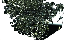 Data gathered at the Diamond Manchester Imaging Branchline (I13-2) have been used to gain a better understanding of lithium battery failure. Lithium-ion batteries provide high power and energy densities, with cells containing pure lithium electrodes theoretically offfering the highest energy storage density by weight and volume. However, lithium metal is unsuitable for commercial use in batteries because it is considered inherently unsafe. Repeated charging and discharging results in electrodeposition of lithium into tree-like dendritic deposits, termed ‘lithium moss’. These deposits continually grow and can short-circuit the battery, leading to battery failure and potentially to fires or explosions.
X-ray Birefringence Imaging
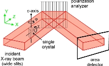 Anisotropic materials are those that exhibit chemical or mechanical differences depending on their orientation. A common example of such a material is wood, where the direction of the grain has an impact on the wood’s characteristics. Birefringent materials are optically anisotropic: the refractive index of the material is dependent on the polarisation state of light passing through it. So, by observing the effect on polarised light passing through a birefringent sample, we can establish the nature of the anisotropy within it. Using the polarising optical microscope, this idea has been used extensively over the last century and has had significant impact on many scientific disciplines including mineralogy, crystallography, materials science, and biological sciences.
Unconventional magnetic order in a 3D Kitaev material
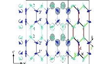 The understanding of magnetism and its effects is important for the fundamental understanding of materials, as well as having the potential to lead to important technological advances. To understand many magnetic effects one needs to study deep at the atomic level, as it is the collective behaviour of all the individual magnetic moments of the constituent atoms that determines the observed macroscopic magnetic properties. At high temperatures magnetic materials are paramagnetic, with the directions of the magnetic moments (spins) randomised by thermal fluctuations. However, at low temperatures magnetic moments tend to point in a particular pattern to minimise the magnetic interactions.
A perfect fit: Understanding how porous organic cages separate rare gases and chiral molecules
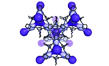 Rare gases exist at very low concentrations in the Earth’s atmosphere; with the exception of argon, noble gases such as xenon and krypton are found at less than two parts per million by volume. It is, however, commercially beneficial to extract such gases due to their value; for example, in commercial lighting, medical imaging and anaesthesia in the case of xenon. In contrast, certain isotopes of these gases can present an environmental hazard. Radon occurs in a naturally radioactive form, which can accumulate in buildings and is a leading cause of lung cancer. It is very difficult to separate such molecules at very low concentrations in the air. It is also difficult to separate chiral molecules in solution, where mirror images of the same molecule have an identical size and shape. Ground-breaking research into porous organic cage molecules has shown selective binding of both noble gases and chiral organic molecules.
B16: Simple inexpensive paper allows advanced X-ray imaging
Hongchang Wang, Sebastien Berujon, Yogesh Kashyap, Julia Herzen, Robert Atwood, David Laundy and Kawal Sawhney

X-ray absorption contrast imaging has proven to be a powerful and invaluable tool since its discovery. However, absorption contrast is weak for samples with weak absorption. In contrast, phase sensitive imaging can allow high contrast with a lower radiation dose than conventional absorption radiography. In addition, X-ray dark-field imaging allows the study of materials in a hitherto unforeseen level. The speckle based phase contrast and dark field contrast imaging has been developed to provide a new imaging method.
Unlike the conventional absorption imaging, both phase and dark field contrast imaging cannot be measured directly. Even though a few imaging techniques have been developed over the last few decades, many of them are limited by either sophisticated experimental conditions or stringent beam properties. We have developed a novel imaging technique to extract the dark field imaging from a stack of speckle images1. The new method can also provide directional dark-field information, which is extremely useful for the study of strongly ordered systems.
In order to reveal the internal structure of soft tissues, the speckle based phase contrast imaging has also been extended from 2D radiography to 3D tomography2. The initial work was carried out on B16, and further experiments were performed on PETRA’s P05 beamline to produce the first 3D image of a human artery by using speckle based technique. As shown in Fig.2, significant enhanced contrast has been observed in the phase contrast computed tomography (CT). The simplicity of the experimental arrangement and speed of measurement gives this new imaging method a distinct advantage over existing X-ray imaging methods, and as such, makes it an attractive technique for in vivo imaging of soft tissue biological systems.

References
1. Wang H. et al. Hard-X-Ray Directional Dark-Field Imaging Using the Speckle Scanning Technique. Phys. Rev. Lett. 114, 103901 (2015).
2. Wang H. X-ray phase contrast tomography by tracking near field speckle. Scientific Reports. 5, 8762 (2015).
I16: Identification of Inversion Domains in KTiOPO4 via Resonant X-ray Diffraction
Federica Fabrizi, Pamela Thomas, Gareth Nisbet and Stephen P. Collins. Submitted to Acta. Cryst. A.
The determination of polar domain patterns is an important prerequisite for investigations of several important phenomena, including magnetoelectric coupling and non-linear optics. For several decades, small polar crystals have been studied effectively using resonant (anomalous) X-ray scattering, where the so-called 'Flack' parameter gives the fraction of each polar orientation. For crystals that are larger than a tiny fraction of a cubic millimetre, this approach fails due to the effects of sample shape and absorption. Moreover, when multiple domains are present it is often essential to observe the detailed domain patterns rather than just the fractional populations.
Here, we demonstrate that by careful choice of a single, highly sensitive Bragg reflection, it is possible to map the surface of an extended crystal with intensity contrast of more than two orders of magnitude, by adopting a pair of photon energies just above and below an absorption edge. Such a map can be carried out with microfocusing optics to achieve micron-scale (or better) spatial resolution. We illustrate this technique with a study of artificially-poled domains in KTiOPO4 1 – a ferroelectric material that exhibits important nonlinear optical properties.
The sensitivity of various Bragg reflections to photon energy, near the Ti K edge, was first investigated numerically using the CCTBX crystallographic library, with Diamond’s DAWN data analysis package as a Python programming platform. These simulations predicted a huge contrast from several reflections, exceeding a factor of 200 for the (417) Bragg reflection.
Measurements were carried out on I16 using the Beamline’s Kirkpatrich- Baez (KB) microfocusing mirror system, giving a spatial resolution, after accounting for beam footprint effects, of around 1.5 x 1.7 microns. The (417) Bragg reflection was measured, with the beam rastered over the sample, for two photon energies, just above and below the Ti K-edge. Thus the ‘domain fraction’ could be determined from the intensity ratio, eliminating any effects of sample inhomogeneity. The resulting map (Fig. 1) shows large regions of up ‘A’ domains towards the top with ‘B’ domains dominating the majority of the scanned area. Towards the centre, a set of artificially generated inversion domains (vertical lines) can be seen, with a periodicity of ~ 9 microns. These were created artificially by applying a high voltage through special electrodes2 in order to improve optical phase-matching for frequency-doubling applications. The results show a very clear map of the artificial domain pattern, and indicated that domain reversal is far from complete, reaching only about 40% at most, with regions where the domain inversion failed completely.
This technique – combining microfocusing with two-energy resonant scattering and simple computer modelling – is likely to find useful application in many important ferroelectric and multiferroic systems. Moreover, the contrast is so high in the present example that a single-energy full-field topographic imaging approach could be applied to studies of domain dynamics in similar systems.
Reference:
1. F Fabrizi et al. Acta. Cryst. A (submitted for publication).
2. T Lyford et al. Acta. Cryst. A (accepted for publication).
Beamline I13 consists of two independently operating branchlines for imaging and coherence. Micro- and nano- structures are imaged in the energy range of 6-35keV, making use of the coherence of light. The beamline covers different scientific areas such as biomedicine, materials science and geophysics.
The imaging branchline, also known as the Diamond Manchester Imaging Branchline, has developed its capabilities further, namely for pink beam imaging. Imaging with micrometre resolution can be performed with millisecond exposure times, full resolution tomography scans are achieved within seconds. The significance of fast tomography has been showcased more recently for the visualisation of the charging/discharging cycle of lithium containing batteries (Eastwood et al. reported in the next section). A better understanding of the dynamics leading to the short circuiting of these systems is an important result applicable to our daily life. Biological studies have been focused on soft tissues and weakly absorbing materials, namely (knee) cartilage, insects and cochlea membranes under realistic conditions.
On the Coherence branch, ptychography (a new imaging technique that promises resolution in the order of 10s of nm or less) is currently implemented as a key technique. More user- friendly software for data acquisition and reconstruction is currently being developed. The method is applied in two dimensions and in combination with tomography, providing high resolution data in three dimensions. For crystalline samples, coherent X-ray diffraction is applied to study stress in nano-crystalline materials. Currently, this research field is being actively pursued by different groups. Photo Correlation Spectroscopy experiments (XPCS) have been performed on colloidal liquids, measuring the transient structure of liquids for the first time.
The instrumentation development has led to major progresses in full-field microscopy and detectors. The microscopy development is enabled through a Diamond wide collaboration on X-ray optics with the group led by C. David at the Paul Scherrer Institute (PSI), Switzerland. A modified scheme for Zernike phase contrast microscopy, which allows resolving 50 nm structures over an extended field of view with minimised artefacts, is currently implemented on the beamline.
Over the past few years, a photon counting detector has been developed in collaboration with the Science and Technology Facilities Council (STFC). EXCALIBUR is a 2kx1.5k pixel detector based on the MediPix3 chip. The detector is mounted on a robot arm. The latter is used in combination with a second robot for moving the detector and a vacuum tube. Both detector and robot arms are about to be made available for the experimental programme.
Beamline I19 continues to support an ever growing user base. Used for chemical crystallography studies, the diffractometer in EH1 will shortly be upgraded to a new instrument. The new diffractometer will have a smaller sphere of confusion, so that a more focused X-ray beam can be used, which will allow smaller sample crystals to be studied. The new diffractometer will be equipped with a Pilatus 2M detector, which will enable significantly faster data collections. The upgrade will occur later this year.
The robotic sample changer in EH1 will become a key element in the new setup and the I19 team has, along with the North East England BAG, developed methodologies for storing a large number of pre-mounted samples under liquid nitrogen and transporting them to Diamond in a Dewar. Once at Diamond the samples are loaded into the robot, while still being held under liquid nitrogen, so that the robot can then mount the samples on to the diffractometer. This procedure is almost identical to that used in macromolecular crystallography (MX) but it is really the first time it has been adopted for small-molecule crystallography at Diamond. This will mean that the robotic sample changer can be used to handle a wider variety of samples and wider use of the robot will significantly improve efficiency for the users.
The EH2 diffractometer is being used for almost all of the sample environment studies. I19 is now using the Pilatus 300K for all experiments, including high-pressure experiments using diamond-anvil cells. The efficiency of the diffractometer has improved dramatically with the introduction of the Pilatus detector and data sets can be routinely collected in under 15 minutes. The Helix open-flow helium cooling device has also been introduced in EH2, making it easy to achieve sample temperatures down to 30 K
The I19 team is continuing to develop peripheral equipment and they have recently introduced a new gas-cell controller in to user operations. The new gascell set up in EH2 increases the accessible pressure range and is straightforward to operate. Offline facilities are being developed to allow users to prepare for gas cell studies in advance of their scheduled beamtime.
Beamline I16 continues to operate very effectively and has recently received more requests for user shifts than at any time during its eight years of operation. The technical focus of the beamline is towards ever-decreasing samples sizes. While there have been no major changes in beamline equipment during the past twelve months, several developments are at the planning stage.
The scientific focus of the beamline remains firmly in condensed matter physics and strongly-correlated electron systems. Specifically, the complex magnetic structures found in 4d and 5d transition metal oxide crystals are of considerable topical interest. Radu Coldea’s group provide an elegant example elsewhere in this Report (Biffin et al), where they describe a complex pattern of counter-rotating moments in g-Li2IrO3. Such a pattern emerges when the Hamiltonian of the interacting spins possesses a strong ‘Kitaev’ term, whereby magnetic coupling is strongly dependent on local bond orientations. Kitaev systems are of intensive theoretical interest as they are exactly solvable, and have been proposed as substrates for future quantum computation.
Materials that spontaneously violate inversion symmetry exhibit a wide range of interesting phenomena such as electric polarisation and magnetoelectric coupling. The ability to map domains in such materials, and correlate them with magnetic structures, is extremely important and one of the main motivations for developing the novel Kirkpatrick-Baez micro-focusing optics for the beamline. With the help of the CCTBX crystallographic software library available at the beamline, a huge sensitivity to polar domain orientation has been demonstrated for specific reflections in KTP, close to the Ti K-edge, with intensity ratios of up to 270. The effectiveness of this technique is illustrated by a micron-resolution study of artificially-poled domain patterns in KTiOPO4 (Fabrizi et al., reported in the next section) – a material of considerable interest for its non-linear optical properties.
The B16 Test Beamline continued its successful operation with the emphasis on a healthy balance between the X-ray optics and detectors development programme and fundamental science research, the latter mainly in the materials science and applied physics areas.
The white-beam compatible submicron-focusing KB (Kirkpatrick-Baez) mirror system installed last year is in routine operation and in constant demand allowing specialised spatially-resolved imaging and scattering/diffraction experiments to be performed on B16 on a few hundreds of nanometres length scale. The channel-cut Si(311) crystal monochromator has been progressively used in the measurements where high angular resolution is required such as topography and reflectivity.
A major technical development that was undertaken this year was the removal of the last 2 m long section of the beamline vacuum pipe which allowed space in the Experimental Hutch for a new Granite block optical table to be fitted. A new micro-tomography station is being installed on this table which will further extend the imaging capabilities of the beamline by allowing experiments with yet unprecedented stability and accuracy to be performed.
Last year, B16 has facilitated the development of a new experimental technique, X-ray Birefringence Imaging (XBI), which follows a close analogy in concept to the polarising optical microscope (Harris et al., reported in the next section). However, rather than polarised visible light, the new technique uses polarised X-rays to interrogate the material of interest, yielding spatially resolved mapping of the X-ray birefringence of the material; which in turn allows the orientational properties of individual molecules and/or bonds within a material to be revealed and visualised.
Another development of novel imaging techniques includes the recent work on speckle-based phase contrast and dark field contrast imaging. In order to reveal the internal structure of soft tissues, the team has also extended the speckle based phase contrast imaging from 2D radiography to 3D tomography (Wang et al., reported in the next section).
Another development of novel imaging techniques includes the recent work on speckle-based phase contrast and dark field contrast imaging. In order to reveal the internal structure of soft tissues, the team has also extended the speckle based phase contrast imaging from 2D radiography to 3D tomography (Wang et al., reported in the next section).

Diamond Light Source is the UK's national synchrotron science facility, located at the Harwell Science and Innovation Campus in Oxfordshire.
Diamond Light Source Ltd
Diamond House
Harwell Science & Innovation Campus
Didcot
Oxfordshire
OX11 0DE
Copyright © Diamond Light Source. Diamond Light Source® and the Diamond logo are registered trademarks of Diamond Light Source Ltd
Registered in England and Wales at Diamond House, Harwell Science and Innovation Campus, Didcot, Oxfordshire, OX11 0DE, United Kingdom. Company number: 4375679. VAT number: 287 461 957. Economic Operators Registration and Identification (EORI) number: GB287461957003.