Bridging the Thickness Gap: Advanced Soft X-ray Imaging Unlocks Magnetic Textures
Aug 19, 2024
Aug 19, 2024
Magnetic materials underpin our high-tech lifestyles and will play key roles in the production of clean energy and next-generation computing technologies. Studying natural magnetic materials can offer insights into animal behaviour, the role of Earth’s magnetic field over time and the formation of the solar system itself. The behaviour of magnetic materials is governed by their underlying magnetisation configuration, including magnetic domains (local areas of uniform magnetisation) and defects such as domain walls.
With magnetic systems varying from the macroscopic scale down to single atoms, and occurring in everything from natural magnetite through to carefully designed topological chiral magnets, researchers require an array of techniques to study them. Exploring magnetic textures on the order of the magnetic exchange length requires spatial resolutions of tens of nanometres or lower.
However, while using a combination of high spatial resolution soft X-ray imaging and electron microscopy allows the analysis of thin samples (≲300 nm) and surfaces, investigation of thicker samples has been limited to hard X-ray dichroic imaging, limiting studies to thin films for most materials, including transition metal magnets.
In work recently published in Physical Review X, an international team of researchers led by Dr Claire Donnelly of the Max Planck Institute for Chemical Physics of Solids has developed a soft X-ray imaging technique for thicker magnets, closing this “thickness gap”. Their work reveals previously inaccessible magnetic textures, and is an exciting development with a range of applications, including the study of naturally occurring magnetic rocks, transition-metal based permanent magnets for energy harvesting, and chiral magnets for spintronics.
Diamond’s soft X-ray ptychography beamline (I08-1) is designed to offer imaging and spectroscopy at resolutions beyond those possible with conventional scanning X-ray microscopes. It works by scanning a beam of partially coherent X-rays across the sample. After recording overlapping diffraction patterns for each position, real-space images can be reconstructed via computational methods. Repeating the process with left and right circularly polarised X-rays allows one to identify differences that correspond to magnetic contrast (X-ray Magnetic Circular Dichroism, or XMCD), which gives a high-resolution image of the magnetic configuration.
Lead author Jeffrey Neethirajan, a PhD student at the Max Planck Institute for Chemical Physics of Solids, said;
In these experiments, we were exploring using ptychography with soft X-rays – low energy X-rays – to probe magnetic materials. Normally, absorption techniques are used to probe magnetic dichroism. However, while we were using spectroscopic methods - seeing how the signal behaves as a function of the X-ray energy - we found that we could use the scattered part of the beam to access the phase dichroism. And that allows us to close the thickness gap and image much thicker magnetic systems with strong dichroic contrast.
The phase dichroism exists for a much wider range of X-ray energies than the absorption dichroism, which generally only occurs in the energy region where the sample is highly absorbing. The X-ray phase dichroism therefore extends soft X-ray magnetic imaging to samples beyond the capabilities of conventional techniques
Benedikt Daurer, Senior Software Scientist at Diamond Light Source, and second author on the paper, continued:
Together with a dichroic measuring approach - collecting ptychographic data with different polarisations – the researchers were able to demonstrate the possibility to obtain magnetic structures from micron-thick samples, substantially widening the range of magnetic samples that can be studied with the I08-1 instrument. We in Diamond’s Data Analysis team are proud to have contributed with our experience in ptychographic reconstructions and related software algorithms.
Dr Donnelly said;
We came to I08-1 specifically to test soft X-ray ptychography for imaging magnetic structure. For our first test run we measured a relatively standard sample, but we were astonished by the quality of the data. It was absolutely beautiful, and allowed us to map this phase dichroism that will not only have huge impact for our research, but also across the field. Working at Diamond, with access to the beamline scientists, and ptychography experts was key, making it possible for us to do really exciting things.
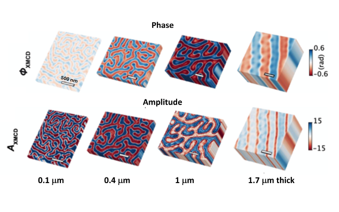
The research team tested this technique with magnetic samples up to 1.7 μm thick, and were able to demonstrate that thick samples of a chiral magnetic material host unconfined states, which opens up exciting prospects for studying knot-like magnetic textures.
Another application lies in palaeomagnetism, the study of magnetic rocks, which offers us clues to the role of Earth’s magnetic field across geological time. Careful examination of the magnetic structure of these particles in relation to the geographical location can tell us how Earth’s magnetic field has changed, and provide insights into the climate when the rocks were formed. It could also be used to study samples from far beyond Earth, which have arrived as meteorites or been collected by spacecraft, to help us understand the conditions present during the formation of the solar system.
Dr Donnelly said;
We’re already working with I08-1 to apply this technique to tomography – 3D imaging – of magnetofossils. We are also very interested in seeing complicated 3D textures present in thicker samples, which could be used for next-gen computing in the next few decades.
Burkhard Kaulich, Principal Beamline Scientist on I08-1 concluded;
Our teams are excited to have contributed to magnetism research that opens new avenues to a broad range of previously inaccessible applications using Diamond’s new I08-1 instrument, in areas such as energy harvesting, spintronics and naturally magnetic materials and structures, like those found in palaeomagnetism. This first highlight demonstrates the potential of the I08-1 instrument, and we must thank Claire and Jeffrey for their invaluable contributions.
To find out more about the I08-1 beamline or discuss potential applications, please contact Principal Beamline Scientist Burkhard Kaulich: [email protected].
Neethirajan JN et al. Soft X-ray phase nano-microscopy of micrometre-thick magnets. Physical Review X 2024. https://doi.org/10.1103/PhysRevX.14.031028

Diamond Light Source is the UK's national synchrotron science facility, located at the Harwell Science and Innovation Campus in Oxfordshire.
Diamond Light Source Ltd
Diamond House
Harwell Science & Innovation Campus
Didcot
Oxfordshire
OX11 0DE
Copyright © Diamond Light Source. Diamond Light Source® and the Diamond logo are registered trademarks of Diamond Light Source Ltd
Registered in England and Wales at Diamond House, Harwell Science and Innovation Campus, Didcot, Oxfordshire, OX11 0DE, United Kingdom. Company number: 4375679. VAT number: 287 461 957. Economic Operators Registration and Identification (EORI) number: GB287461957003.