Access denied: Stopping rabies virus from entering cells
Sep 28, 2022
Sep 28, 2022
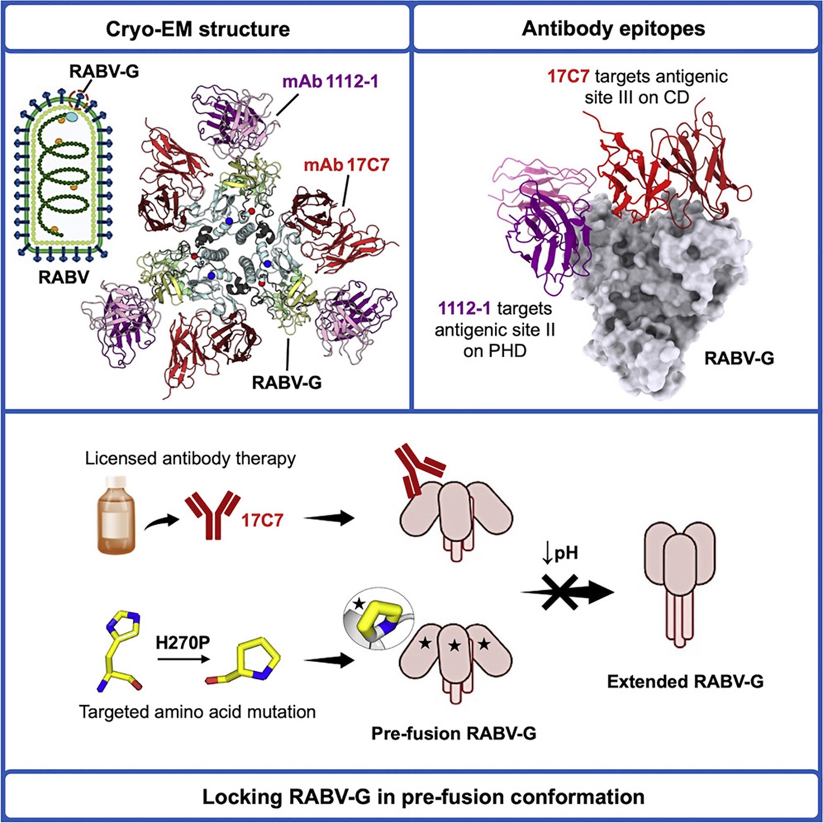
Rabies virus can cause fatal neurological disease when access to medication is scarce. Tackling this issue will require novel vaccines and therapies to be developed and the viral surface protein may be a suitable target for these interventions. This protein arranges in triplets, but the structure of this arrangement has not yet been determined and the interaction between this triplet and therapeutic agents remains to be characterised. In a recent publication in Cell Host & Microbe, a research team at the University of Oxford collaborated with the electron Bio-Imaging Centre (eBIC) at Diamond Light Source to solve the cryo-electron microscopy (cryo-EM) structure of the triplet in complex with two therapeutic agents. Building on this work, the scientists investigated the mechanism by which licensed therapies inhibit the viral protein. The structural insight gained from this work will allow for strategic development of new vaccines and therapies against the virus.
Rabid animals typically spread rabies virus to humans by biting and this can cause a lethal neurological infection. Despite the development of vaccines and therapeutics, over 60,000 people die each year from rabies. Most of these deaths occur in African and Southeast Asian countries, where the virus is endemic in animals and access to medical intervention is insufficient. There is a growing necessity to design accessible therapeutics in response. Currently, vaccines can prevent rabies, but multiple doses are often required to achieve protection. Exposed individuals can additionally be treated with antibodies — components of the immune system that detect and block specific microorganisms (e.g. bacteria or viruses). There is concern however that currently available antibodies may not be protective against new variants of the virus and additional antibodies need to be produced.
Vaccines and antibody therapies target a single protein on the surface of the rabies virus. This aptly named ‘fusion’ protein engages with cellular proteins and triggers fusion of the virus with the cell, allowing infection to begin. Fusion proteins come together in triplets and change shape to drive fusion. The triplets initially form an open tripod (i.e. in which the three ‘legs’ are splayed). To allow fusion, the legs then fold into a closed-tripod arrangement. The researchers purified the fusion protein as a triplet in the open-tripod arrangement to study its architecture and association with antibodies.
The team purified the fusion proteins in the open-tripod arrangement by introducing mutations that stabilised the structure. This alone is an important step towards efficiently purifying the fusion triplets for novel vaccine development. The researchers subsequently prepared the fusion triplets in complex with two antibodies, both of which have been shown to offer protection but only one of which is licensed for treatment so far. To determine the structure of the whole complex, the team visited the electron Bio-Imaging Centre (eBIC) at Diamond where they used a technique optimised for determining the structure of large biological complexes, known as cryo-electron microscopy (cryo-EM). By virtue of the efficiency of the cryo-EM facilities at eBIC, over 12,000 images were taken, with more than three million complexes being captured, and the structure of the fusion tripod and bound antibodies was refined to near-atomic detail.
Dr Weng M Ng, one of the lead authors of the study, recalls his experience at eBIC:
The beamline scientists provided valuable help and made remote data collection smooth and straightforward during the COVID-19 lockdowns. The Titan Krios microscope we used allowed us to collect high-quality images.
The structure of individual fusion proteins had previously been determined but not in the biologically relevant tripod arrangement until now. The structure revealed regions of the fusion protein triplet that may trigger the change in shape required for fusion as well as regions that are bound by the protective antibodies. These regions have been identified as suitable targets for the development of new therapeutic antibodies.
The mechanism by which the licensed antibody blocks the fusion protein was not known. The antibody may obstruct any engagement between the fusion proteins and cell proteins. Alternatively, it may prevent the fusion triplets from switching from an open-tripod to a closed-tripod arrangement. The switch normally occurs under acidic conditions. However, the switch at low pH was inhibited when the fusion triplet was bound by the licensed antibody, revealing that the antibody locked the fusion protein in the open-tripod arrangement that is unable to trigger fusion.
The structure of the complex highlights how the fusion protein triplet can be stabilised for vaccine development, how the tripod structure may change shape, and how the fusion triplet interacts with protective antibodies.
Professor Alexander Douglas, the corresponding author of the study, remarks on the implications of this work:
Rabies prevention requires multiple vaccine doses, but these treatments are often prohibitively expensive or out of reach in low-income countries where fatality from the disease is high. Structure-guided vaccinology could fast-track development of vaccines that can be delivered to remote areas in need.
To find out more about eBIC or discuss potential applications, please contact Principal Electron Microscopist Daniel Clare: [email protected]
Ng W. M. et al. Structure of trimeric pre-fusion rabies virus glycoprotein in complex with two protective antibodies. Cell Host & Microbe 30 (Sep 2022).
DOI: 10.1016/j.chom.2022.07.014.

Diamond Light Source is the UK's national synchrotron science facility, located at the Harwell Science and Innovation Campus in Oxfordshire.
Diamond Light Source Ltd
Diamond House
Harwell Science & Innovation Campus
Didcot
Oxfordshire
OX11 0DE
Copyright © Diamond Light Source. Diamond Light Source® and the Diamond logo are registered trademarks of Diamond Light Source Ltd
Registered in England and Wales at Diamond House, Harwell Science and Innovation Campus, Didcot, Oxfordshire, OX11 0DE, United Kingdom. Company number: 4375679. VAT number: 287 461 957. Economic Operators Registration and Identification (EORI) number: GB287461957003.