The dynamic duo
Aug 3, 2020
Aug 3, 2020
A novel, user-friendly platform for looking inside cells in three-dimensions has been developed at B24 at Diamond Light Source. The combination of super-resolution structured illumination microscopy and soft X-ray tomography, detailed in a publication in Cell, was put to the test by tracking the early stages of reovirus infection.
The Biological Cryo-Imaging beamline, B24 at Diamond, comprises the transmission cryo-soft X-ray tomography (cryo-SXT) microscope, which allows users to visualise biological samples under near-physiological conditions. Recently, the beamline has been enhanced with the addition of a cryo-structured illumination microscope (cryo-SIM).
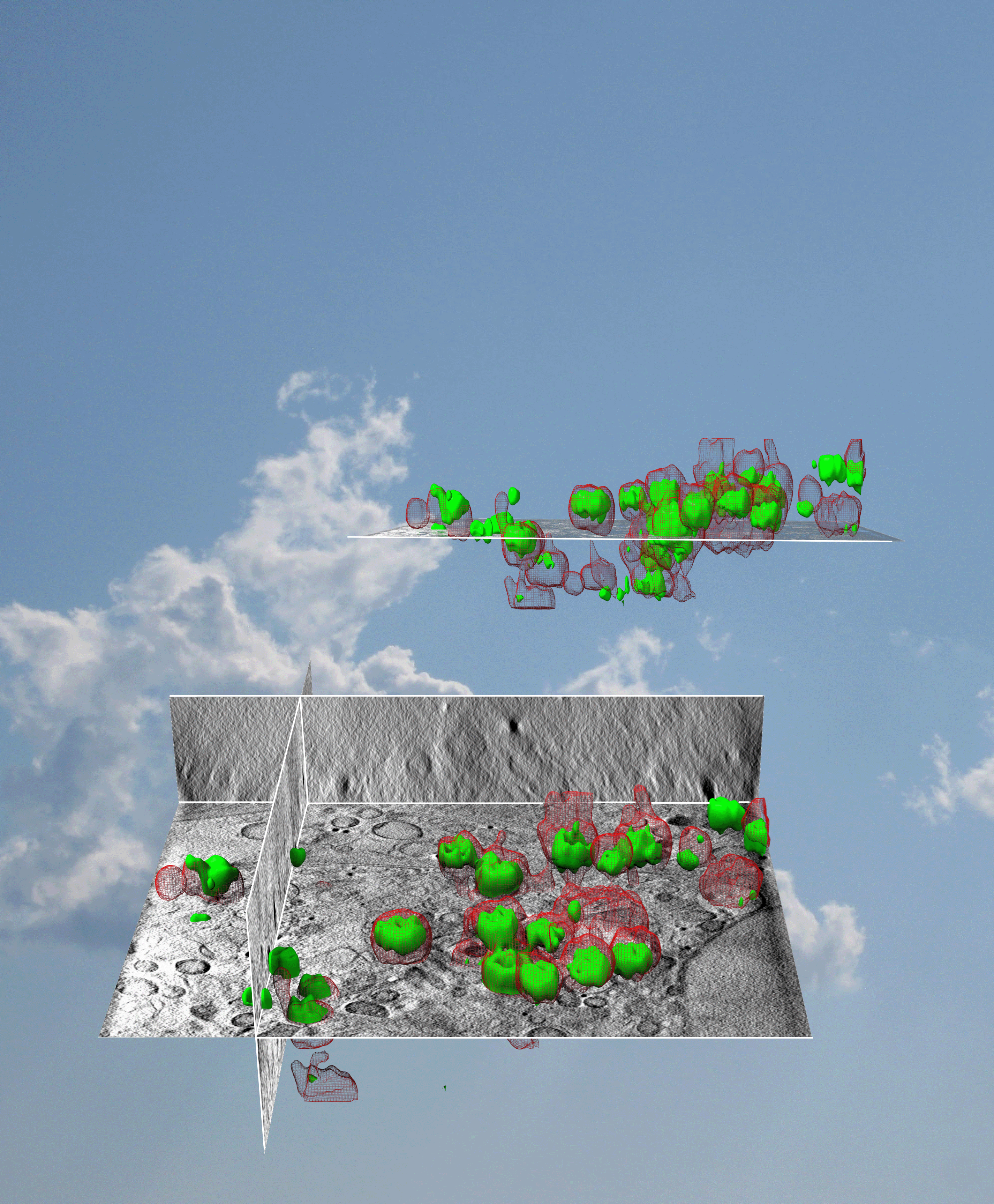
A collaborative effort of European scientists saw these two techniques merged to allow users to combine the ultrastructural data from SXT with the fluorescence data from SIM. The power of this novel imaging platform was demonstrated by studying reovirus. The two high-resolution imaging techniques allowed data to be superimposed on the same cell samples in 3D to track events during the early stages of infection.
Gaining high-quality imaging data from cells is a challenge; not only immobilising the cells in a way that does not introduce artefacts, but also ensuring the imaging techniques yield information that can cross-correlate.
At B24, the development of a new imaging platform has filled this unmet need. The power of the cryo-SIM and cryo-SXT microscopes have been combined to give valuable insights into the inner workings of biological material. This work was the result of a fruitful collaboration between Diamond and research groups and facilities across Europe (University of Oxford, UK, Heidelberg University Hospital, Germany, Université de Nantes, France, CryoCapCell, Paris, France and Micron, which is based at the Department of Biochemistry at the University of Oxford).
Dr Maria Harkiolaki, Principal Beamline Scientist at B24 and lead author of the study, explained the motivation behind combining the techniques,
One method gives you a chemical signature and the other method gives you ultrastructure and content, literally a CT scan of a cell with highlighted areas of interest. Put them together and you not only have a 3D map of a cell, but also the location within that map of any chemical entity you are interested in.
The new imaging platform was put through its paces by tracking the way in which reoviruses infect a cell. Reoviruses are well-studied benign viruses and promising candidates for gene therapy vectors. Although they been researched extensively, the way in which the viruses enter cells and release their contents is debated. The virus uses endosomal vesicles to enter cells, but it is not fully understood how it escapes this vesicle to produce progeny.
Human cells grown on gold grids were infected with reovirus and samples were plunge-frozen after 1, 2 and 4 hours. At each of these time points the cells were imaged using SIM-SXT to determine the location of the virus and its carrier vesicles.
Dr Harkiolaki explained;
When you look at cells in a normal microscope, they aren’t very photogenic – you don’t see many things as they are usually transparent. But when we use these lasers, we can highlight and track the virus and we had a reporter chemical that told us when the carriers’ membranes had been compromised
After 2 hours, the fluorescence data showed that the virus had already escaped from the vesicles that contained it. The data was enhanced by merging this information with the X-ray microscopy, which allowed the team to see both carriers and virus alongside other cellular structures. The vesicles still appeared round in the images despite the virus already escaping, which hinted towards a gentle exit via pores within the vesicle because preservation of the round shape would not have been possible if the virus had ruptured the vesicle to escape.
The elucidation of the infection mechanism of the reovirus was made possible with the new platform, and its applications extend far beyond this proof-of-concept experiment. It has already been used to track cell morphology changes during development, delivery of anti-cancer compounds, pathogen clearance by immune cells and seeing into yeast, algae, bacteria and archaea.
Importantly, this work perfectly demonstrates what can be achieved by multidisciplinary collaboration. Dr Harkiolaki said;
We have virologists from German and UK institutes with the real-life need for multidimensional imaging of cell populations at near physiological states, imaging development experts from Diamond and the University of Oxford and finally software developers from Diamond and France coming together to produce a really integrated imaging platform that is accessible and user-friendly.
The platform will continue to be improved and will also be fully automated in the future. In the meantime, despite the COVID-19 pandemic, experiments can be conducted remotely and the methodology is accessible online to help the scientific community take advantage of developments at B24.
To find out more about B24, or to discuss potential applications, please contact Principal Beamline Scientist Maria Harkiolaki: [email protected].
3D Correlative Cryo-Structured Illumination Fluorescence and Soft X-ray Microscopy Elucidates Reovirus Intracellular Release Pathway, ScienceDirect, 23 July 2020.
DOI: 10.1016/j.cell.2020.05.051.

Diamond Light Source is the UK's national synchrotron science facility, located at the Harwell Science and Innovation Campus in Oxfordshire.
Diamond Light Source Ltd
Diamond House
Harwell Science & Innovation Campus
Didcot
Oxfordshire
OX11 0DE
Copyright © Diamond Light Source. Diamond Light Source® and the Diamond logo are registered trademarks of Diamond Light Source Ltd
Registered in England and Wales at Diamond House, Harwell Science and Innovation Campus, Didcot, Oxfordshire, OX11 0DE, United Kingdom. Company number: 4375679. VAT number: 287 461 957. Economic Operators Registration and Identification (EORI) number: GB287461957003.