Cryo-EM structures show how vertical single β-barrel viruses manage self-assembly
Jul 24, 2019
Jul 24, 2019
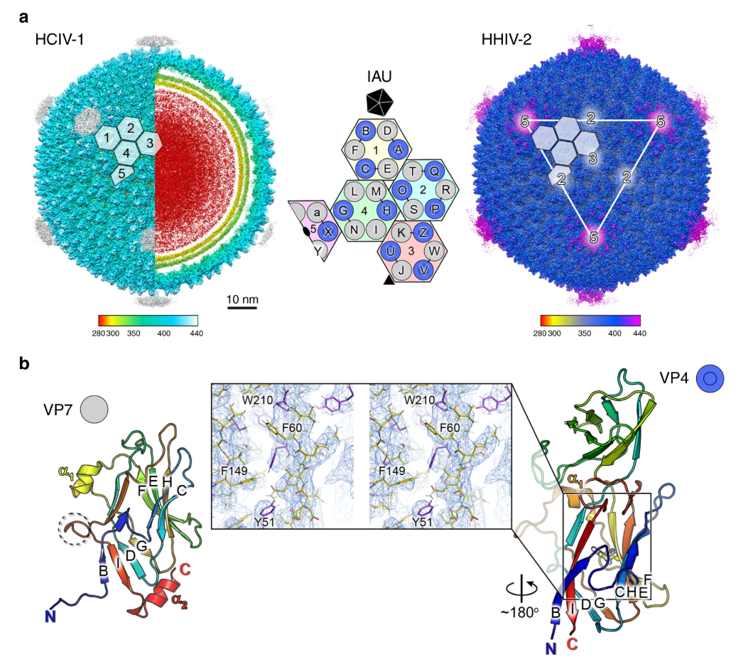
In 1977, Carl Woese et al introduced the three-domain system of biological classification that divides life forms into Archaea, Bacteria and Eukaryotes. This was the first time that the differences between Archaea and Bacteria were recognised. The first observed archaea were extremophiles, able to survive and thrive in extremely harsh conditions, but ongoing research has found them in a wide range of habitats; they are particularly abundant in the oceans. Archaea form part of the microbiota of all organisms, including humans, and their unique survival capabilities mean they can be used to enhance our understanding of early life on Earth. We are just beginning to investigate viruses that can infect Archaea, and in work recently published in Nature Communications, an international team of researchers determined the near-atomic structures of HCIV-1 and HHIV-2 by cryo-electron microscopy (cryo-EM). Their results show how nature has solved the complexity of arranging vertical single β-barrels and provide insights into the evolutionary consequences of the fusion event of the two consecutive major capsid protein (MCP) genes, which has ultimately led to the membrane-less vertical double β-barrel MCP assembly seen in adenovirus.
Professor Nicola Abrescia at the CIC bioGUNE (Spain) and his colleagues Prof Bamford and Dr Oksanen at the University of Helsinki enjoy poking around in salt pans and other unexplored environmental niches, combining ‘fishing trips’ to collect new viruses with work or leisure trips around the world. This work investigates two related viruses, membrane-containing Haloarcula californiae icosahedral virus 1 (HCIV-1) and membrane-containing Haloarcula hispanica icosahedral virus 2 (HHIV-2), samples of which were collected in Thailand and Italy, respectively. Initial research to identify the host and virus and the conditions for virus propagation and purification highlighted an unexpected complexity in the arrangement of the proteins forming the capsid shell.
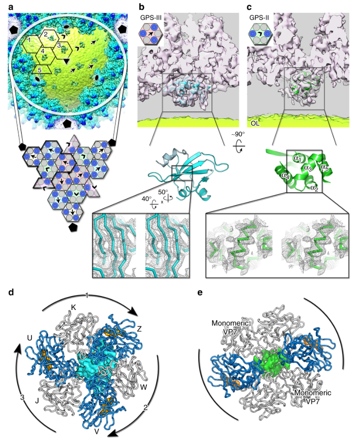
Together with proteomics data, structures determined by cryo-EM based provide insights into the assembly mechanism of this type of virus, and into those with membrane-less double β-barrel MCPs. As Prof Abrescia explains,
Two proteins located between the capsid shell and the outer membrane leaflet operate as a kind of GPS. These two different proteins act as navigation buoys for the direction of assembly.
The results explored in this paper add to our understanding of how gene duplication and gene fusion events can lead to a decrease in assembly complexity and allow us to test, in computational biology, how this apparent assembly simplification leads to the encoding of more complex information in the fused MCP gene.
To find out more about the electron Bio-Imaging Centre (eBIC), or to discuss potential applications, please contact Director of eBIC, Peijun Zhang: [email protected]
Santos-Pérez I et al. Structural basis for assembly of vertical single β-barrel viruses. Nature Communications 10, 1184 (2019). DOI:10.1038/s41467-019-08927-2.

Diamond Light Source is the UK's national synchrotron science facility, located at the Harwell Science and Innovation Campus in Oxfordshire.
Diamond Light Source Ltd
Diamond House
Harwell Science & Innovation Campus
Didcot
Oxfordshire
OX11 0DE
Copyright © Diamond Light Source. Diamond Light Source® and the Diamond logo are registered trademarks of Diamond Light Source Ltd
Registered in England and Wales at Diamond House, Harwell Science and Innovation Campus, Didcot, Oxfordshire, OX11 0DE, United Kingdom. Company number: 4375679. VAT number: 287 461 957. Economic Operators Registration and Identification (EORI) number: GB287461957003.