Non-destructive X-ray diffraction
Jul 28, 2017
Jul 28, 2017
X-ray diffraction (XRD) is an analytical method used to determine the nature of crystalline materials, through measurements of crystal unit dimensions. Although XRD is ubiquitous, especially in fields such as geology, materials science, and biology, to obtain successful results, the analyte material must be finely ground and homogenised. Experiments conducted at Diamond Light Source, however, have demonstrated the efficacy of a novel, non-destructive XRD technique that does not require the standard sample preparations. The high-resolution back-reflection energy-dispersive XRD (EDXRD) method has previously been proposed, but the experimental campaign conducted on the Core X-ray Absorption Spectroscopy beamline (B18) is the first time that this technique has been used to obtain high-fidelity crystallographic and structural measurements. The results from several geologic specimens, fossils, and archaeological artefacts—as recently reported in Acta Crystallographica A—indicate that the back-reflection EDXRD method is insensitive to sample shape and to the sample–beam distance. It is thus suitable for characterising rare and valuable samples, such as extra-terrestrial rock and meteorite materials, or objects of cultural importance.
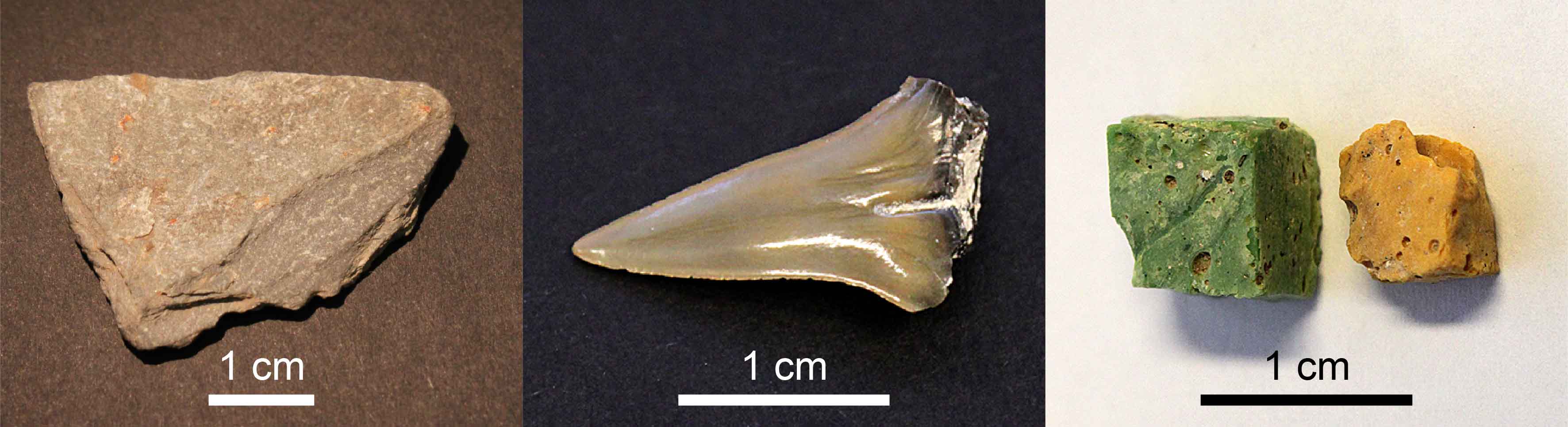
Figure 1: Photographs of the mica schist (left), fossil shark tooth (centre), and Roman mosaic tesserae (right) specimens used for the experimental campaign.
Limitations of X-ray diffraction
XRD has long been part of the standard arsenal for chemical analyses of crystalline substances. Nonetheless, the geometrical requirements of standard (i.e. Bragg–Brentano) XRD mean there are strong constraints on sample positioning and morphology. In general, it is necessary to grind samples to a fine powder to ensure a large number of crystallites are exposed to the beam and to produce a completely flat sample surface. This destructive preparation, however, is undesirable for valuable specimens or those that are available in very small quantities.
Alternative XRD geometries have previously been proposed to offer relaxed morphology and positioning constraints. For example, transmission XRD approaches (including tomographic energy-dispersive diffraction imaging) can be used to obtain the phase composition of samples1–3. These methods, however, generally require high-energy X-ray (i.e. synchrotron) sources and there is an upper limit to the depth of the samples that can be analysed. In addition, parallel-beam XRD (in which a parallel beam of X-rays is used in a reflection-mode geometry) offers a significant degree of insensitivity to sample morphology and positioning4. This approach is now widely used in modern laboratories, but as an angle-dispersive XRD approach, it tends to suffer from shadowing problems at low diffraction angles.
Experimental validations
The new study, led by Dr Graeme Hansford, Instrument Scientist at the University of Leicester’s Space Research Centre, sees the experimental validation of a concept he first conceived, back in 2010 when he was brainstorming an XRD instrument that would require no sample preparations or specific positioning (e.g. for use on an asteroid). Since then, Dr Hansford has used theoretical and ray-trace modelling methods to demonstrate the validity of the back-reflection geometry EDXRD technique5, and has experimentally tested it in low-resolution configurations6. The key criterion is to use an X-ray diffraction scattering angle (2θ) as close to 180° as feasible. The primary aim of the Diamond experiments, therefore, was to prove the back-reflection EDXRD technique remains insensitive to sample morphology and detection geometry in a high-resolution setup.
For the experimental campaign, Dr Hansford—together with Dr Stuart Turner (University of Leicester), Professor Andrew Shortland (Centre for Archaeological and Forensic Analysis, Cranfield University), and Professor Patrick Degryse (Centre for Archaeological Science, University of Leuven, Belgium)—used the Core X-ray Absorption Spectroscopy beamline (B18) at Diamond. This beamline is typically used for general-purpose X-ray absorption spectroscopy (with a highly focused beam), but in this case an unusual configuration was implemented. For the back-reflection EDXRD technique to work properly “the beam needs to illuminate as many crystallites as possible”, Dr Hansford explained, “we therefore chose to defocus the beam and increase its footprint on the sample”. The team also used a Si(111) monochromator to achieve their high-resolution spectral measurements and a silicon drift detector for X-ray detection.
Several well-understood specimens were selected for the high-resolution back-reflection EDXRD tests (see Fig. 1). These included geologic samples (e.g. mica schist and chert), pressed-powder pellets (single minerals and complex assemblages), fossils, and archaeological objects. The scientists found that the results were insensitive to the shape and texture of a sample’s surface. In addition, moving the sample by up to 16 mm (in the sample–beam direction) made essentially no difference to the spectra. The team also successfully characterised unprepared geological samples and identified the mineral phase responsible for the colour of a Roman mosaic tessera.
Practical applications
Having demonstrated that the high-resolution back-reflection EDXRD technique can be used to make effective crystal phase and structure identifications, Dr Hansford’s team are now aiming to develop the methodology for smaller-scale laboratory or museum settings. Without the need for time-consuming, difficult, and destructive sample preparation procedures, the new methodology is particularly suited for the analysis of precious specimens, e.g. for characterising extra-terrestrial rock samples. As highlighted by Prof Shortland, this technique also has the potential “to answer new questions about our most valuable and interesting historical and archaeological objects”, such as their provenance and authenticity.
To find out more about the B18 beamline, or to discuss potential applications, please contact Principal Beamline Scientist Dr Giannantonio Cibin: [email protected]
Hansford GM et al. High-resolution X-ray diffraction with no sample preparation. Acta Crystallographica A 73, 293–311 (2017). DOI:10.1107/S2053273317008592.
1. Cernik RJ et al. X-ray colour imaging. J. R. Soc. Interface 5, 477–481 (2008).
2. Lazzari O et al. Reconstructive colour X-ray diffraction imaging – a novel TEDDI imaging method. Analyst 134, 1802–1807 (2009).
3. Scarlett NVY et al. Energy-dispersive diffraction studies of inert anodes. J. Appl. Cryst. 42, 502–512 (2009).
4. He BB. Two-Dimensional X-ray Diffraction. Wiley (2009).
5. Hansford GM. Back-reflection energy-dispersive X-ray diffraction: a novel diffraction technique with almost complete insensitivity to sample morphology. J. Appl. Cryst. 44, 514–525 (2011).
6. Hansford GM. X-ray diffraction without sample preparation: proof-of-principle experiments. Nucl. Instrum. Methods Phys. Res. A 728, 102–106 (2013).

Diamond Light Source is the UK's national synchrotron science facility, located at the Harwell Science and Innovation Campus in Oxfordshire.
Diamond Light Source Ltd
Diamond House
Harwell Science & Innovation Campus
Didcot
Oxfordshire
OX11 0DE
Copyright © Diamond Light Source. Diamond Light Source® and the Diamond logo are registered trademarks of Diamond Light Source Ltd
Registered in England and Wales at Diamond House, Harwell Science and Innovation Campus, Didcot, Oxfordshire, OX11 0DE, United Kingdom. Company number: 4375679. VAT number: 287 461 957. Economic Operators Registration and Identification (EORI) number: GB287461957003.