World first photothermal nanospectroscopy at Diamond
Mar 1, 2016
Mar 1, 2016
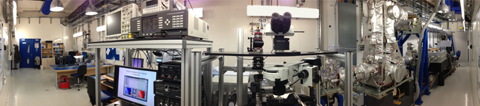
Panorama of the MIRIAM beamline, with the AFM-IR setup at the centre.
Infrared (IR) microspectroscopy is a quantitative analytical and non-destructive method valuable in a wide variety of research, from medical biochemistry and histology, to the science of new and composite materials, and the physical chemistry of surfaces. It is also used for studies in cultural heritage and archeology, biomineralogy, and geology. In biological and medical studies this technique is particularly relevant since it is routinely used to pinpoint single cell biology (e.g. responses to test drugs for cancer therapy or stem cell differentiation/control by chemical molecules), but the diffraction limit means that it is unable to clearly resolve the sub-cellular structure below the micron scale.
Dr Gianfelice Cinque (Principal Beamline Scientst for MIRIAM) and his team have been investigating coupling IR spectroscopy with the use of photothermal nanoprobes to produce higher resolution data. The advantages of using IR synchrotron radiation (IR-SR) as the light source, over benchtop sources/lasers, is that the bright, broadband radiation enhances the sample vibrational spectra quality on small samples, and dramatically speeds up data acquisition while exploring the whole IR range.
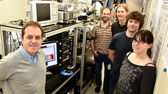 Gianfelice Cinque (left), Principal Beamline Scientist for MIRIAM, with some members of the research team (l-r): Mark Frogley, Paul Donaldson, Chris Kelley, and Ann Fitzpatrick.
Photothermal probes
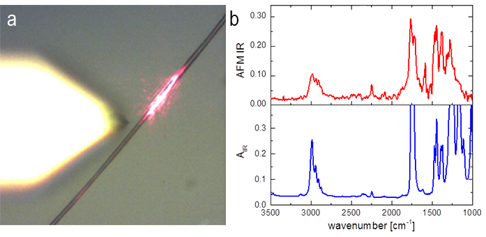
An ultrafast mechanical chopper spinning in vacuum at circa 20,000 rpm has been implemented on MIRIAM to modulate the illumination beam and set up to the timescale necessary to maintain the excitation locally at the sub-micron level - a technical challenge in its own right. Tuning this chopper to work in resonance with the AFM cantilever produces a larger oscillation and a larger signal. When you correlate the wavelength to the oscillation amplitude you’ve got an absorption spectrum and can tell which molecular species (IR fingerprint) are present quantitatively (absorption intensity).
Dr Cinque says, “the two real advantages of using synchrotron radiation in our field are brightness and broadband. Brightness is how many photons per second per unit area and angle can be focused effectively onto the smallest optical spot at the microscope. If you want to look at a small object, you need to focus as many photons as possible onto the sample spot. The broadband SR advantage means that you’ve got all the wavelengths at the same time, and this is not common when using lasers, for example. So brightness and broadband are what synchrotrons bring to diffraction-limited IR microspectroscopy, or eventually nanospectroscopy like we just proved."
Diamond’s IR radiation spans the largest possible range, extending from the near-IR up to the far-IR (or THz) region. And the beam is 100-1000 times brighter in the mid-far-IR than any conventional broadband IR source.

Diamond Light Source is the UK's national synchrotron science facility, located at the Harwell Science and Innovation Campus in Oxfordshire.
Diamond Light Source Ltd
Diamond House
Harwell Science & Innovation Campus
Didcot
Oxfordshire
OX11 0DE
Copyright © Diamond Light Source. Diamond Light Source® and the Diamond logo are registered trademarks of Diamond Light Source Ltd
Registered in England and Wales at Diamond House, Harwell Science and Innovation Campus, Didcot, Oxfordshire, OX11 0DE, United Kingdom. Company number: 4375679. VAT number: 287 461 957. Economic Operators Registration and Identification (EORI) number: GB287461957003.