Improving corneal surgery
Jul 27, 2011
Jul 27, 2011
The cornea is the external lens of the eye, responsible for refracting incoming light onto the crystalline lens behind, which in turn focuses it on to the retina. It also plays a protective role, shielding the rest of the eye from dust and infection. To function, the cornea must be transparent to visible light, possess high mechanical strength, and have precisely defined curvature to focus.
90% of the thickness of the cornea is made of the stroma, a thick transparent layer primarily composed of collagen fibrils. The transparency stems from the small, uniform diameter and regular separation of these fibrils, whereas mechanical strength is related to their arrangement in the tissue. The shape of the cornea is almost spherical at the centre of the eye (near the visual axis), flattening out towards the white sclera. The shape of the cornea is thought to be strongly influenced by the arrangement of collagen fibrils in the stroma and surrounding tissues, and this knowledge is key to understanding how corneal disease and surgery can affect vision.
Professor Keith Meek, Dr Craig Boote, and a team from the Cardiff University have been using Diamond’s I22 beamline and ID13 at the ESRF in Grenoble, France, to study the microstructure of the peripheral cornea and the limbus, where the cornea meets the sclera. This work has been published in the Biophysical Journal.
Human corneas without any history of corneal disease or surgery were obtained from the Bristol Eye Bank. Microfocus Wide Angle X-ray Scattering (WAXS) on ID13 was used to examine the preferred fibril orientation and the relative number of fibrils oriented in a particular direction. The SAXS experiments at Diamond used a conventional (rather than microfocus) beam, enabling greater sampling and reducing the signal to noise ratio, enabling the team to accurately measure fibril size. The results showed that the fibril diameter is constant in the central 6mm of the cornea but increases rapidly when moving out towards the periphery.
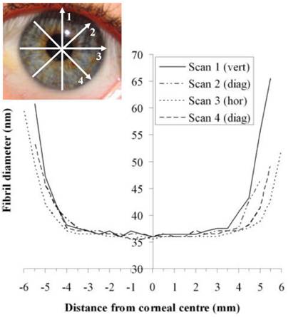 |
| Figure 1: SAXS experiments on beamline I22 have determined how collagen fibril diameter varies across the human cornea. |
“Our study showed that the integration of collagen fibrils between the cornea and limbus is not circularly symmetrical, but varies noticeably depending on direction. This may be an important factor in understanding how the cornea responds to surgery involving peripheral incision – for example, cataract surgery. Astigmatism is a common complication following such surgery and our research will help to explain observations by clinicians that the extent of astigmatic changes depends on the location the cut is made.”Prof Keith Meek, University of Cardiff
Dr Burghammer, beamline scientist at ESRF and co-author on the paper, says, “The investigations on cornea illustrate very well the efficient use of complementary techniques across beamlines at different synchrotron radiation sources. The combined application of scanning micro-diffraction and small angle scattering for biomedical applications is expected to be a rapidly growing field in the years to come.”
Quantification of Collagen Organization in the Peripheral Human Cornea at Micron-Scale Resolution, Craig Boote, Christina Kamma-Lorger, Sally Hayes, Jonathan Harris, Manfred Burghammer, Jennifer Hiller, Nicholas Terrill, Keith Meek, Biophysical Journal 101, July 2011.
DOI: 10.1016/j.bpj.2011.05.029

Diamond Light Source is the UK's national synchrotron science facility, located at the Harwell Science and Innovation Campus in Oxfordshire.
Diamond Light Source Ltd
Diamond House
Harwell Science & Innovation Campus
Didcot
Oxfordshire
OX11 0DE
Copyright © Diamond Light Source. Diamond Light Source® and the Diamond logo are registered trademarks of Diamond Light Source Ltd
Registered in England and Wales at Diamond House, Harwell Science and Innovation Campus, Didcot, Oxfordshire, OX11 0DE, United Kingdom. Company number: 4375679. VAT number: 287 461 957. Economic Operators Registration and Identification (EORI) number: GB287461957003.