Synchrotron applications such as coherent X-ray diffraction and X-ray photon-correlation spectroscopy require detectors with a very small pixel size. Furthermore, the detector should have a high frame rate, large dynamic range, high detection efficiency and be also radiation hard. The Medipix range of readout chips (Medipix2 and Medipix3), with a pixel pitch of 55 µm, emerged as good candidates to develop a large area detector for the aforementioned applications. However, reducing the pixel size leads to an increase in the charge-sharing effect, which affects the detection efficiency and energy resolution of the detector. In order to overcome this effect, characterization of different hybrid photon-counting detector configurations were performed on beamline B16 at Diamond Light Source. A planar and a columnar 3D silicon sensor, each one bump-bonded to the Medipix2 readout chip, were two of the studied assemblies. The other assembly consisted of a planar silicon sensor bump-bonded to the Medipix3 readout chip. The results of radiation hardness and charge-sharing suppression obtained with the Medipix3 configuration encouraged the development of a new large area detector based on the Medipix3 readout chip.
X-ray hybrid photon-counting detectors are becoming a standard component in many synchrotron beam lines around the world since they offer several advantages over traditional X-ray detection technologies. They offer high frame rate, large dynamic range, high count-rate capability and high signal-to-noise ratio. They are used for macromolecular crystallography, small-angle X-ray scattering, surface and interface diffraction and X-ray reflectivity experiments. However, for applications such as coherent X-ray diffraction and X-ray photon-correlation spectroscopy, the detector pixel size required should be the order of 50 µm. At the moment, only the Medipix range of readout chips achieves this requirement, with their 55 µm pixel size.
Reducing the pixel size leads to an increase in charge-shared events between boundary pixels and, therefore, to a degradation of the energy resolution and detection efficiency of the detector. Charge-shared events occur when an X-ray impinges close to the pixel borders and the generated charge-cloud splits across the boundary pixels. Then the record of the event by the pixel is strongly dependent on the energy threshold discriminator set, causing count losses or double counts by a high or low energy threshold discriminator setting respectively. When working with monochromatic beams, the charge-sharing effect across pixel borders can be minimized (but not completely suppressed) by setting the energy threshold discriminator at half the impinging energy.
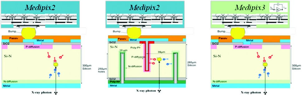
Figure 1: Characterized assemblies. Left: Planar silicon sensor with conventional readout mode, referred to as standard assembly. Center: Columnar 3D sensor bump-bounded to Medipix2. Right: Planar silicon sensor bump-bonded to a Medipix3.
Three different detector assemblies were studied (Fig. 1). The first was defined as the standard assembly and consisted of a planar silicon sensor bump-bonded to a Medipix chip operated in conventional readout mode, which was either the conventional readout configuration for Medipix2 or the Single Pixel Mode readout configuration for Medipix3. Independently of the Medipix version, both readout chips consists of an array of 256 x 256 pixels of 55 µm size each. The other two detector assemblies were characterized and compared to the standard assembly. Hybrid photon-counting detectors consist of two parts: a photodiode sensor and a CMOS readout chip. Each of the other two assemblies under study approached the charge-sharing effect and radiation sensitivity problem by using the latest developments of each field.
The experiments to characterize these detectors were performed on beamline B16 at Diamond Light Source. Their uniformity was studied by scanning a cluster of nine adjacent pixels in steps of 2.5 µm with a 15 keV micro-focused X-ray beam of 2.2 µm × 2.9 µm size. The energy threshold discriminator was set at half the incoming energy to minimize the charge-sharing effect. Even with this threshold setting, the charge-sharing effect cannot be completely eliminated for the standard assembly. The charge-cloud generated close to the corners of a pixel was only partially recorded as shown in Figure 2, with only a detection efficiency of 13% in some spots.
One of the detector assemblies is based on 3D silicon pixel array sensors, which is the latest design in silicon photodiode sensors. Whereas a planar silicon sensor consists of planar electrodes (see Fig. 1), a 3D silicon sensor consists of a three dimensional array of p and n electrodes that penetrate into the detector bulk perpendicular to the surface.1 They are of interest for correcting the charge-sharing effect since their electric field pattern causes the charge carriers to drift horizontally far from the pixel boundaries. Studies of the charge-sharing effect with a 3D silicon sensor bump-bonded to a Medipix2 readout chip are discussed in.2 Although an improvement in the charge-sharing effect was observed with 3D silicon sensors, an additional count-loss due to the electrodes was observed as is shown in Figure 2.
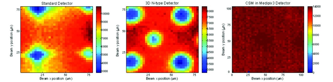
Figure 2: Detection efficiency results of each detector assembly after scanning a cluster of nine pixels with the micro-focussed beam. Left: Standard assembly. Center: Columnar 3D sensor bump-bonded to Medipix2. Right: Planar silicon sensor bump-bonded to a Medipix3.
The other detector assembly is based on the latest developments of CMOS technology. It consists of a planar silicon sensor bump-bonded to a Medipix3 readout chip.3 Medipix3 is the latest generation of photon-counting readout chips of the Medipix family. It is implemented in 0.13 µm CMOS technology which leads to an increase in the functionality associated with each pixel over Medipix2. One of the new operational modes implemented in the front-end architecture is the Charge Summing Mode (CSM). This mode consists of a charge reconstruction and hit allocation algorithm which eliminates event-by-event the low energy counts produced by charge-shared events between adjacent pixels. Results showed4 a uniform detection efficiency when the detector was operated in CSM mode with all detected events within a 4% standard deviation. There were no count losses or double counts (Fig. 2), which corroborated the good performance of this new operating mode at eliminating charge-shared events between boundary pixels.
Additionally, the Medipix3 design is expected to be more radiation hard that its predecessor Medipix2. After the positive results on eliminating the charge-shared effect with the Medipix3-based detector, its global response to radiation hardness was evaluated by exposing the detector to the white beam produced by the bending magnet at B16. In this study5 a Medipix3 detector area of 5 mm diameter was exposed to very high levels of X-ray radiation. Results showed that radiation damage effects started at much higher radiation dose levels than when using a Medipix2 chip. The study also showed a recovery after being exposed to 1.8 MGy with a large dose rate of 3.5 kGy/s. The recovery of the detector took place without applying any special conditions to it within a period of 25 days.
The positive results obtained for the Medipix3-based detector on charge-shared events suppression and radiation hardness encouraged the development of a large area detector based on Medipix3 readout chip for synchrotron applications which require high image quality.
E.N. Gimenez, R. Ballabriga, M. Campbell, I. Horswell, X. Llopart, J. Marchal, K.J.S. Sawhney, N. Tartoni, D. Turecek, Characterization of Medipix3 with Synchrotron Radiation, IEEE Trans. Nucl. Sci., vol. 58, pp. 323-332 (2011).
E.N. Gimenez, R. Ballabriga, M. Campbell, I. Horswell, I. Dolbnya, X. Llopart, J. Marchal, K.J.S. Sawhney, N. Tartoni, D. Turecek, Evaluation of the Radiation Hardness and Charge Summing Mode of a Medipix3-based detector with Synchrotron Radiation Nuclear Science Symposium Conference Record IEEE (November 2010)
References
- Pellegrini, G. et al., “First double-sided 3-D detectors fabricated at CNM-IMB”, Nucl. Instr. and Meth. A. 38, 592 (2008).
- Gimenez, E.N. et al., “3D Medipix2 detector characterization with a micro-focused X-ray beam”. Nucl. Inst. Meth. A (2010). doi:10.1016/j.nima.2010.06.140
- Ballabriga, R. et al. “The Medipix3 Prototype, a Pixel Readout Chip Working in Single Photon Counting Mode with Improved Spectrometric Performance”. IEEE Trans. on Nucl. Sci. Vol. 54, 1824-1829 (2007).
- Gimenez, E.N. et al., “Study of charge-sharing in MEDIPIX3 using a micro-focused synchrotron beam”, Journal of Instrumentation 2011_JINST_6_C01031 (2011), doi: 10.1088/1748-0221/6/01/C01031
- Gimenez, E.N. et al. “Evaluation of the Radiation Hardness and Charge Summing Mode of a Medipix3-based detector with Synchrotron”. IEEE Nuclear Science Symposium Conference Proceeding. J. Inst. (Nov 2010).