The experiment undertaken on I16 showed strong evidence of twinning, from the ‘speckled’ Bragg peaks of manganites crystallites. This was consistent with a new theory of the connection between ab-twinning, a classic disorder mode of these layered materials, and the phase of the domain that contributes to a given Bragg peak.
The very slight difference in a and c lattice constants found in orthorhombic Cuprate and Manganite crystals allows domains to match along their 101 and 10-1 directions. These materials can show “ac twinning” in which small domains alternate between one orientation and the other on the nanometre scale. When such a structure is illuminated with a coherent beam focussed down to 2 microns, as we achieved at I16, the Bragg peaks are broken up into speckles. We demonstrated this effect on specially grown crystals of composition PrCaMnO, abbreviated PCMO. It is straightforward to show that the number of speckles in the Bragg peak is equal to the number of domains in the beam. We could therefore estimate the twin domain size to be around 50-100 nm.
Our new understanding of the origin of this speckle shows that the visibility is proportional to the phase shift between ac and ca domains, which is itself simply related to the order of diffraction and the magnitude of the lattice constant difference. Higher angle peaks and those in particular lattice directions have more visibility than others. Our method represents an entirely new way of measuring the a-c lattice constant difference and allows sensitive testing of models of disorder in crystals.

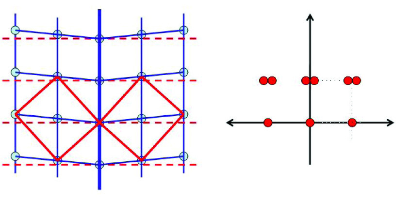
Figure 1: Twin boundary between two orthorhombically-distorted domains of PCMO (left), illustrating the splitting of the Bragg peaks (right).
The crystals of PCMO was found to exhibit the characteristic splitting of their Bragg peaks, as expected for the rotated domains obtained by matching the diagonals of the ac and ca unit cells with each other. This is shown schematically in Fig. 1. A splitting of 0.25 degrees was observed for some reflections and not others, again as expected from the crystallography. Sometimes the splitting was in the direction of momentum transfer; sometimes seen directly on the detector.

Figure 2: Coherent Diffraction pattern of a PCMO crystal pair showing speckles extending between two centres of a split Bragg peak due to twinning. [PCMO3-7, frame 28]
Since the beam was made coherent during the experiment by use of a coherence-defining aperture and then focussed down to 2 x 2 microns to get enough intensity from the micron-sized crystals, the speckles and fringes of the diffraction patterns of both split Bragg peaks were found to interfere with each other and give a single merged coherent diffraction pattern as shown in Fig. 2.
Our new model of the shearing of the lattices giving rise to a phase structure is illustrated in Fig. 3. The coloured stripes represent the (real-space) phase within the bicrystal, shown as a translucent 3D box. The phase ramps positively on both the left and right sides of the bicrystal boundary in the centre. The calculated diffraction pattern in Fig.3 (left) shows the splitting of the peak due to the phase structure and the intermodulation of the diffraction in between.


Figure 3: (a. left) Colours represent the phase model of the bicrystal, shown as a translucent 3D box. (b. above) calculated diffraction pattern . [n7-ramp045-iso]
References
[1] I. K. Robinson and R. Harder, Coherent Diffraction Imaging of Strains on the Nanoscale, Nature Materials, 8, 291-298 (2009).
[2] S. J. Leake, M. C. Newton, R.Harder, and I. K. Robinson, Longitudinal Coherence Function in X-ray Imaging of Crystals, Optics Express,17, 15853 (2009).
[3] M. C. Newton, S. J. Leake, R. Harder and I. K. Robinson, Three-Dimensional Imaging of Strain in a Single ZnO Nanorod, Nature Materials, 9, 120-124 (2010).
Principal Publications and Authors
M.A.G. Aranda, F. Berenguer, R.J. Bean,X. Shi,G. Xiong, S.P. Collins, C. Nave, I.K. Robinson. Journal of Sychrotron Radiation accepted.
Funding Acknowledgement
European Research Council advanced grant.
Diamond Light Source is the UK's national synchrotron science facility, located at the Harwell Science and Innovation Campus in Oxfordshire.
Copyright © 2022 Diamond Light Source
Diamond Light Source Ltd
Diamond House
Harwell Science & Innovation Campus
Didcot
Oxfordshire
OX11 0DE
Diamond Light Source® and the Diamond logo are registered trademarks of Diamond Light Source Ltd
Registered in England and Wales at Diamond House, Harwell Science and Innovation Campus, Didcot, Oxfordshire, OX11 0DE, United Kingdom. Company number: 4375679. VAT number: 287 461 957. Economic Operators Registration and Identification (EORI) number: GB287461957003.