
___________________________________
Industrial Liaison Group:
Tel: +44 (0) 1235 778797
E-mail: [email protected]
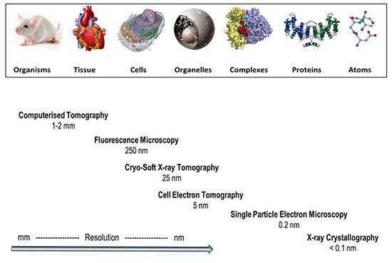
Over the last few decades, we have seen a growing interest in the use of high-resolution imaging to study the structures of biological samples. Increased access to synchrotron sources has also driven the development of imaging capabilities and widening use of cryo-soft X-ray tomography across life sciences.
A recent study at Diamond looks at the life-cycle of malaria, discovering a new step in the stages of infection.
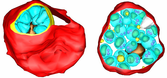
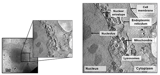
X-rays pass through a biological sample and get attenuated based on carbon content (carbon-rich biological structures absorb soft x-rays preferentially.) The generated projections are recorded at small angular increments to create a tilt series of images that provide structural information at different orientations. These are then reconstructed into tomograms, the cellular equivalent of CT scans used in hospitals, to produce a 3D image. Other techniques can be used, in correlation with tomography, to zoom in on specific areas of interest and really interrogate the data.
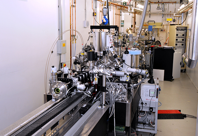 For cryo X-ray tomography to take place at Diamond, samples are grown on flat grids coated with a carbon substrate to stabilise the cells. Fiducial markers are included in the sample to allow accurate and automated reconstructions of the data. The samples are then frozen using liquid nitrogen-cooled liquid ethane and preliminary mapping undertaken to ensure samples are suitable for imaging (for example, they have not been contaminated during plunge freezing and they retain their original form and structure). X-rays are used to generate mosaic images which provide overviews of whole regions and specific areas of interest are identified for data collection.
 The amount of data generated from X-ray imaging is vast and processing is fully automated, allowing for near real-time data analysis; in-house alignment and reconstruction software is used to pull together the 3D image. Thereafter, a process of segmentation enables the user to classify objects or areas of interest. Once cellular features have been segmented and classified, further analysis can be done to look at size, shape and localisation to gain further biological insight.
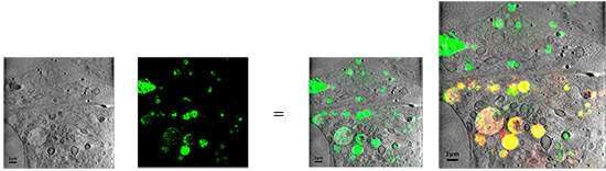
Cryo soft X-ray tomography is still an emerging technique for the area of life sciences and on the whole is still only available using synchrotron sources. Over recent years, it has grown in use due to improved accessibility and improved capabilities. It is a very effective technique to visualise the entire structure of cells in a non-destructive way, enabling them to observe whole cells in a near native state. In addition, data can be correlated across imaging techniques.
Cryo-soft X-ray tomography: using soft X-rays to explore the ultrastructure of whole cells
Maria Harkiolaki , Michele C. Darrow , Matthew C. Spink , Ewelina Kosior , Kyle Dent , Elizabeth Duke
Emerging Topics In Life Sciences; DOI: 10.1042/ETLS20170086

Diamond Light Source is the UK's national synchrotron science facility, located at the Harwell Science and Innovation Campus in Oxfordshire.
Copyright © 2022 Diamond Light Source
Diamond Light Source Ltd
Diamond House
Harwell Science & Innovation Campus
Didcot
Oxfordshire
OX11 0DE
Diamond Light Source® and the Diamond logo are registered trademarks of Diamond Light Source Ltd
Registered in England and Wales at Diamond House, Harwell Science and Innovation Campus, Didcot, Oxfordshire, OX11 0DE, United Kingdom. Company number: 4375679. VAT number: 287 461 957. Economic Operators Registration and Identification (EORI) number: GB287461957003.

 Industrial Liaison Office