Diamond peers inside cells hosting Chlamydiae bacteria
Sep 22, 2021
Sep 22, 2021
A powerful new X-ray imaging technique at Diamond Light Source has helped researchers shed light on an important class of bacteria responsible for a range of diseases in humans and animals.
Best known as the cause of a sexually transmitted infection, Chlamydiae are a diverse group of pathogens whose strains can also lead to pneumonia and blindness.
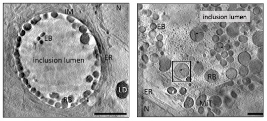
Chlamydiae form communities inside the cells of their hosts in compartments known as inclusions. During their life cycle they transition between two forms, responsible for replication within a host cell and infection of neighbouring cells respectively.
But little is known about how this infection system works in practice, or how the nature of Chlamydiae communities influences the individual bacteria members within them. Such knowledge could open up new treatment avenues for the diseases caused by these pathogens.
The senior author of this new study, which uses cutting-edge cryo-Soft X-ray Tomography (cryo-SXT) at the correlative cryo-imaging beamline B24 at Diamond to investigate this pathogen, is Dr Maud Dumoux. Dr Dumoux, Technology Lead for cryo-imaging at the Rosalind Franklin Institute, said:
Chlamydiae are unique bacteria, as they grow only inside cells. Most strains form a specialised compartment in which hundreds – sometimes thousands – of bacteria multiply and differentiate themselves between non-infectious and infectious forms, preparing for release into the wider environment to infect other cells in the body. How communities of Chlamydiae organise these events, and how each community has an impact on its individual bacteria, are intriguing questions that have implications beyond this particular pathogen.
In this paper, the research team – from the Franklin, Diamond Light Source, Research Complex at Harwell, and Oxford University – sought to uncover how communities of Chlamydiae bacteria regulate and organise their space, and how basic factors such as concentration and size of bacteria can impact the life cycle. To do this, the researchers used the correlative cryo-imaging capacity of beamline B24 and specifically, the transmission X-ray microscope for cryo-SXT data collection.
Dr Maria Harkiolaki, Principal Beamline Scientist at B24, explains further:
The techniques we use allow us to inspect delicate structures in cells and find out how things are organised inside them. Cryo-SXT data collection at this beamline is done through a rapid user-friendly process that captures intra-cellular behaviour and interactions quickly and in intricate detail. B24 is one of only four facilities in the world that provides this technology.
Commenting on their results, Dr Dumoux said:
Surprisingly, we found that concentration of bacteria is not correlated with differentiation into the infectious form. In other words, at the time we observed the infected cells, a high concentration did not trigger preparation for exiting the cell and infecting other cells. However, we did show that higher concentrations lead to smaller individual bacteria, whereas bacteria given space can reach very large volumes – similar to the apocryphal idea that fish will grow to the size of their pond. This is very interesting because it demonstrates, as is often the case in life science, that things are not black and white. Here, there are shades of grey as the bacteria adapt or respond to their environmental pressures. It also opens up new questions about whether different types of bacteria have different roles within the community.
The researchers conclude that each ‘inclusion’ (a specialised compartment of bacteria within an infected human cell) operates as an autonomous community that influences the characteristics of individual bacteria within it, and that bacterial concentration is a key factor in determining those characteristics. With cryo-SXT now established as a useful and rapid technique for studying Chlamydiae, future research will shed light on the evolution of infection and communication between bacterial communities, which could open up new therapeutic opportunities.
To find out more about Diamond’s correlative cryo-Imaging beamline for the life sciences (B24), or to discuss potential applications, please contact the Principal Beamline Scientist Maria Harkiolaki: [email protected]
Patrick Phillips et al. Single Cell Cryo-Soft X-ray Tomography Shows That Each Chlamydia Trachomatis Inclusion Is a Unique Community of Bacteria. Life (2021). DOI: 10.3390/life11080842

Diamond Light Source is the UK's national synchrotron science facility, located at the Harwell Science and Innovation Campus in Oxfordshire.
Diamond Light Source Ltd
Diamond House
Harwell Science & Innovation Campus
Didcot
Oxfordshire
OX11 0DE
Copyright © Diamond Light Source. Diamond Light Source® and the Diamond logo are registered trademarks of Diamond Light Source Ltd
Registered in England and Wales at Diamond House, Harwell Science and Innovation Campus, Didcot, Oxfordshire, OX11 0DE, United Kingdom. Company number: 4375679. VAT number: 287 461 957. Economic Operators Registration and Identification (EORI) number: GB287461957003.