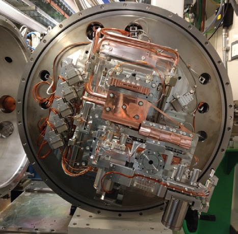
- Figure 1: The double crystal monochromator upgrades for I03, I04 and VMXi.
Academic and industrial researchers from around the globe use Diamond’s macromolecular crystallography (MX) beamlines to reveal and explore the three dimensional shape of large biological molecules. An intimate knowledge of shape and the arrangement of chemical properties in, for example, proteins, DNA and viruses, provides functional understanding of these important biological assemblies. This leads to better interpretation of the processes of life undertaken by cellular pathways and complex assemblies enabling discoveries spanning from fundamental biology through to MX becoming an integrated part of the drug discovery pipeline.
Highlights
I02 - Dynamic pores in the HIV capsid import nucleotides for DNA synthesis
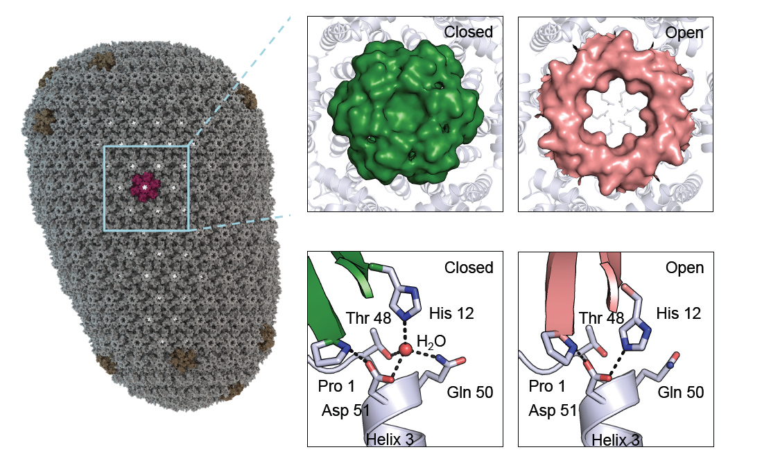 HIV is a retrovirus that needs to copy its RNA content into DNA within a host cell. How it avoids being detected and destroyed by our immune system during this process is currently unknown, and fully understanding this mechanism is paramount to developing an effective HIV vaccine.
Read more about this I02 highlight.
I03/I04/I24 - Bacterial outer membrane assembly machines
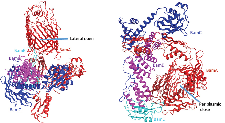 Gram-negative bacteria, mitochondria and chloroplasts all have outer membranes, which contain a variety of proteins. As well as helping to pull together the membrane components, outer membrane proteins have an important role in importing nutrients, exporting waste products and transporting proteins. In Gram-negative bacteria, outer membrane proteins are inserted and folded into the outer membrane by the β-barrel assembly machinery (BAM), but the mechanism of this process is poorly understood. BAM itself is a complex that comprises five subunits: one outer membrane protein called BamA and four lipoproteins (fat-loving proteins that are fixed to the membrane) known as BamB, Bam C, BamD and BamE.
Read more about this MX highlight.
I04 - Identification of a novel binding site on the glucagon receptor
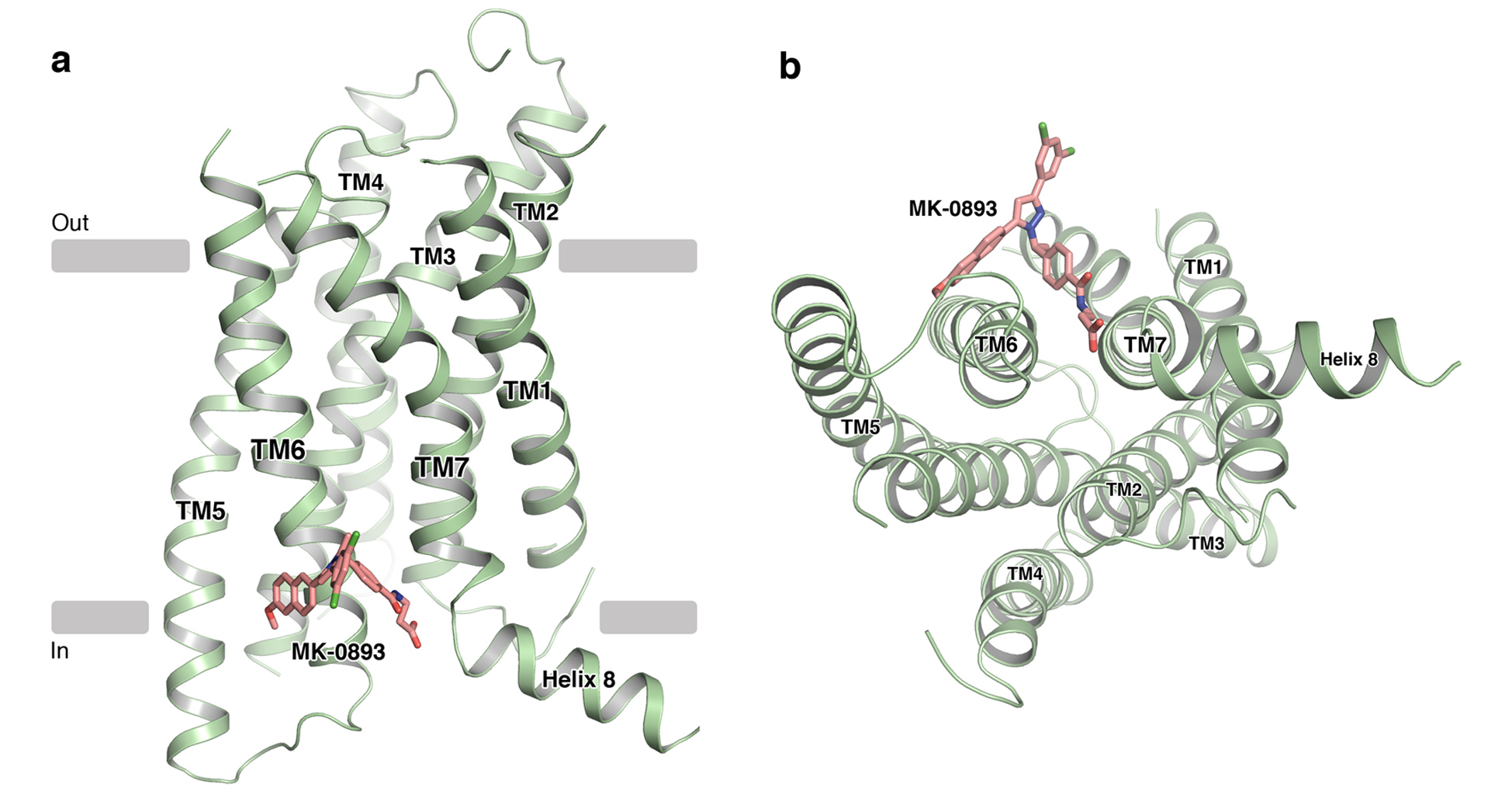 Glucagon is a key hormone made by the pancreas, which raises blood sugar levels by acting on cell surface receptors. The interaction between glucagon and the glucagon receptor (GCGR) is therefore an important area of research for the treatment of diabetes. The inhibition of glucagon signalling has been proposed as a method to reduce glucose production in the liver, but there is little understanding of the mechanism of action of recently developed small molecules that have been designed to target the GCGR. The aim of this study was to solve the crystal structure of one such inhibitor bound to the GCGR.
Read more about this MX highlight.
I02/ I03/ I04 and I04-1 - Insights into broad-spectrum antivirals
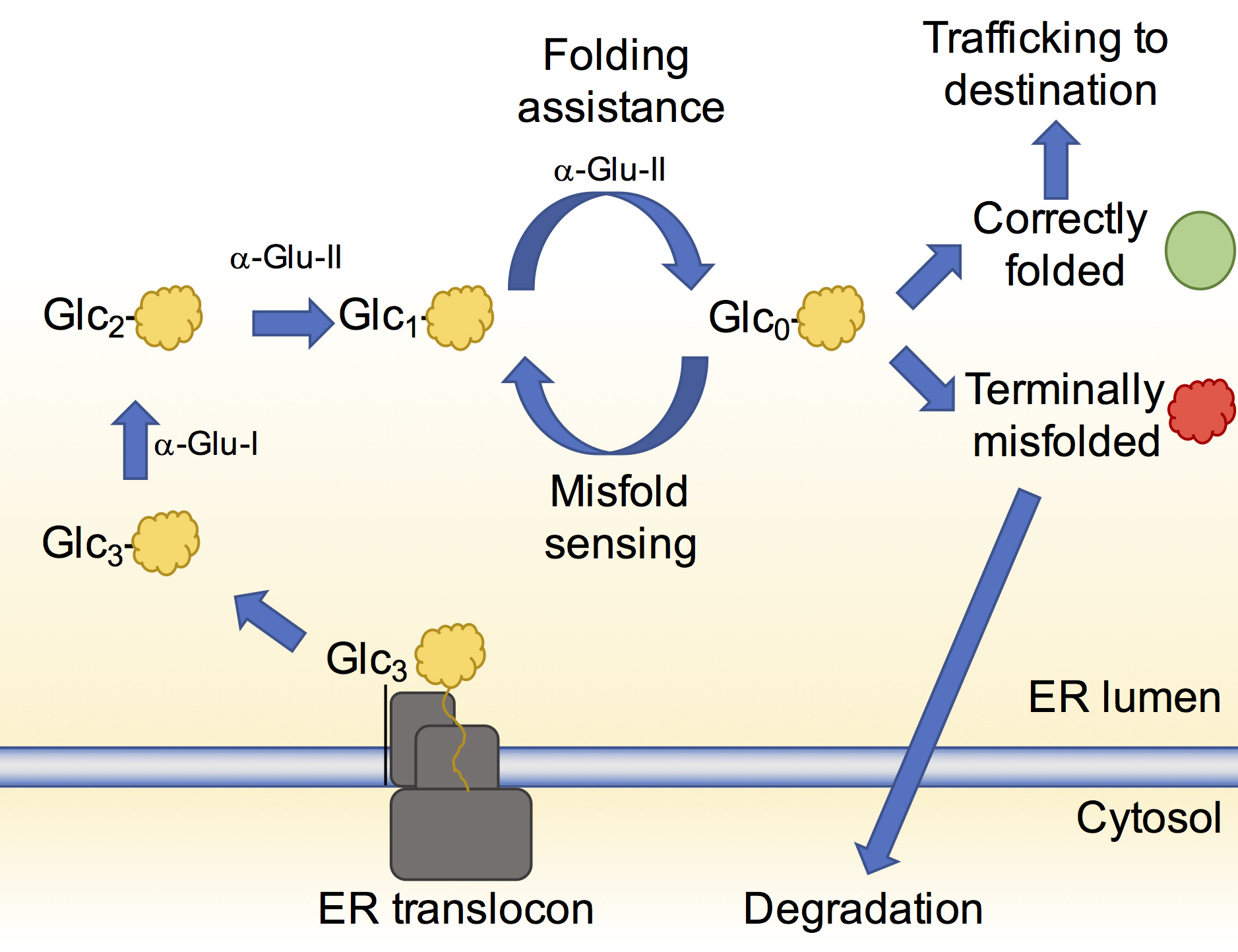 Broad-spectrum antibiotics are designed to combat a wide range of bacteria; however, the same therapeutic approach cannot be applied to viruses. Currently, viral infections need to be treated individually with specific antiviral drugs, but a novel therapeutic strategy has been developed to inhibit a shared viral enzyme known as α-glucosidase II. This enzyme is responsible for folding viral surface glycoproteins to ensure the viral envelope is properly constructed during replication.
Read more about this MX highlight.
I23 - Using the unique capabilities of the long-wavelength Diamond beamline to solve a protein structure
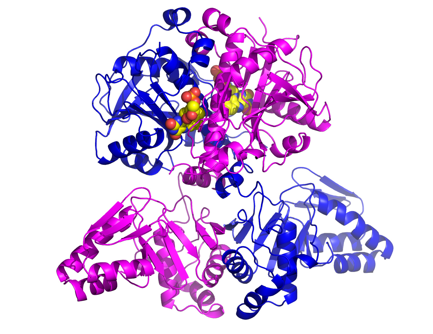 The structure of an oxidase protein from a key cyanobacteria pathway has been uncovered for the first time, using the new Long-Wavelength Macromolecular Crystallography beamline (I23) at Diamond Light Source. This is the first novel structure to be solved on the I23 beamline using the sulfur single-wavelength anomalous diffraction (S-SAD) phasing method.
Read more about this MX highlight.
I24/B21 - The oncogenic G-protein coupled receptor Smoothened is regulated by its extracellular domain
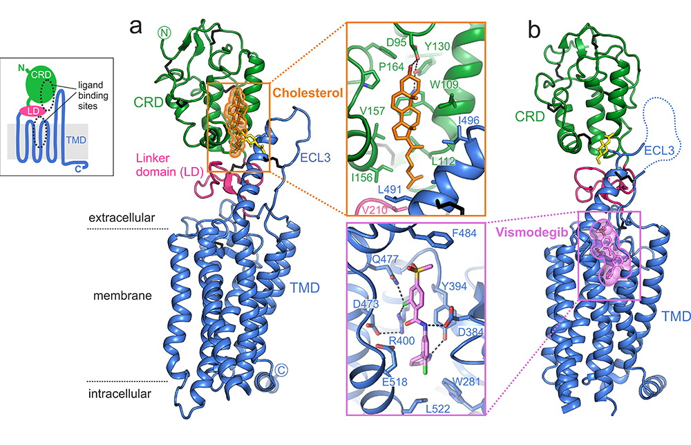 The Hedgehog signalling pathway is one of the many ways cells communicate with each other, and it is involved in embryonic development, cancer genesis and stem cell function. The structure of a protein which helps transmit these signals across a cell membrane has been solved using the tuneable Microfocus beamline (I24) and the High Throughput Small Angle X-ray Scattering (SAXS) (B21) beamline at Diamond Light Source. The protein, Smoothened, is a G-protein-coupled receptor (GPCR), spanning the cell membrane and protruding beyond it. Researchers used I24 to generate diffraction data from small crystals (no larger than 10-20 μm long) of the protein, and B21 to understand the dynamics and large-scale domain reorientations of the protein in solution.
Read more about this MX highlight.

Diamond Light Source is the UK's national synchrotron science facility, located at the Harwell Science and Innovation Campus in Oxfordshire.
Diamond Light Source Ltd
Diamond House
Harwell Science & Innovation Campus
Didcot
Oxfordshire
OX11 0DE
Copyright © Diamond Light Source. Diamond Light Source® and the Diamond logo are registered trademarks of Diamond Light Source Ltd
Registered in England and Wales at Diamond House, Harwell Science and Innovation Campus, Didcot, Oxfordshire, OX11 0DE, United Kingdom. Company number: 4375679. VAT number: 287 461 957. Economic Operators Registration and Identification (EORI) number: GB287461957003.