
___________________________________
Industrial Liaison Group:
Tel: +44 (0) 1235 778797
E-mail: [email protected]
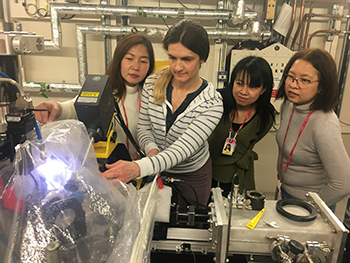
 In this article we speak with Dr Worawikunya Kiatponglarp who is a beamline scientist at SLRI, the national synchrotron of Thailand.
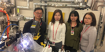
Along with four colleagues and the support of the Industrial Liaison team, she was able to complete a study at Diamond, through the Newton Fund programme. The work was commissioned by Pan Rajdhevee Group Public Co. Ltd with a focus on their commercial sunscreen product, Minus.
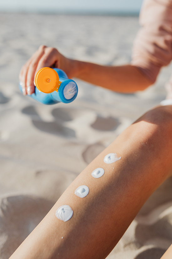 As people have started to become more aware of the dangers of ultra violet rays, the rise in popularity of sunscreen has been immense. Many of the products currently available use Titanium dioxide (TiO2) and Zinc oxide (ZnO) nanoparticles.
Formulas with these nanoparticles are able to block both UVA and UVB radiation and prevent skin irritation. Unlike traditional sunscreens, these nanoparticles are more transparent, less viscous and more easily absorbed; they also leave no unsightly white film on the skin.
Despite their obvious benefits, many people have raised concerns about their ability to penetrate deep into the skin below the stratum corneum (SC) and into the bloodstream. If this were possible, it could result in oxidative stress, inflammation, allergies, and even cancer.
Working with SLRI scientists, Pan Rajdhevee Group Public Co. Ltd (a well-known cosmeceutical company in Thailand), we wanted to investigate the extent to which their new product, Minus-Sun Facial Sun Protection SPF50+ PA+++ penetrated into the skin.
With help from scientists at the Synchrotron Light Research Institute (SLRI) in Thailand and Diamond Light Source, we were able to track the nanoparticles from the cream using a tiny beam of X-rays to determine how deep they were penetrating into the different layers of the skin.
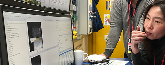
 Using I18 microfocus beamline at Diamond the team studied very thin skin samples - treated with the topically applied sunscreen - for 2, 6, and 12 hrs to see how deep the TiO2 and ZnO nanoparticles (NPs) had penetrated into the skin.
The skin was then cross-sectioned into very thin samples and micro-XRF/XAS experiments conducted. These techniques gave us insight into the local distribution of NPs and their oxidation states in each layer.
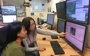
We used the micro-XRF beamline at SLRI, Thailand to study the distribution of Ti and Zn in a cross-section of skin samples. However, the study was limited to the epidermis layer due to the restricted capabilities of the beamlines - spot size (30 µm) and photon flux. From these results we found that these two elements had accumulated as clusters.
To probe into the deeper layers required a much smaller beam size and higher intensity X-rays. Thanks to Diamond’s microfocus beamline, (1 micron beam), the team were able to map a very tiny spot where Zn or Ti clusters had accumulated. This allowed us to see in which sub-layers the NPs were most likely to accumulate.
 The results of the study demonstrated that TiO2 and ZnO NPs in Minus commercial sunscreen were able to penetrate and accumulate in the SC, stratum lucidum and upper St. granulosum of the epidermis layer but not into the deeper epidermis layer and dermis layer at ppm scale. The team noted that some zinc was detected in the hair follicle and tissue, but this was of a different species than the one in the sunscreen product.
The findings of this study will be of significance importance for future applications of topical nanoparticle-based technology strategies, in terms of the dose and dermal distribution of the particles. Synchrotron-XRF and XAS techniques have shown their potential to measure elemental distribution in skincare products.
When we started this research project using the synchrotron facility in Thailand, it was a challenge for us to be able to detect such low concentrations of nanoparticle within a very thin biological sample. It turned out that we could only detect areas where there was a high level of nanoparticle accumulation, specifically on the epidermis layer. By using I18 beamline at Diamond, we were able to see even more detail of accumulated nanoparticles in each sub-layer of the epidermis.
The Newton Fund provides a great opportunity for researchers from developing countries where advanced equipment such as a synchrotron facility is difficult to access. It allowed us not only to solve the industrial research challenges we were experiencing but also for our beamline scientists to be trained at Diamond’s advanced light source facility, with the support of top scientists.
Diamond is well placed to support these types of studies. They have advanced equipment, and highly experienced scientists to provide advice and support for your research. Through the Newton Fund, groups are supported both technically and financially.
We have learned so much, for example, how to handle fragile samples, and good sample preparation techniques (which then lead to good data sets). When dealing with biosamples, it’s essential to get every step right. Sometimes, different tools can make a difference; sometimes, a particular skill is required. Apart from using advanced equipment and working with highly skilled scientists, we have really loved working with the team at Diamond, and spending time with them both socially and professionally.
From my personal experience, I had a wonderful and productive time at Diamond. Not only did I get lots of useful data, but I was truly inspired by the scientific community at Diamond. I would highly recommended other scientists to apply for the Newton Fund and collaborate with Diamond.
This study clearly shows that micro-XRF/XAS is effective at examining accumulated nanoparticles, in cosmetic products, within the skin layers. This method has the potential for wider use within the product development cycle and could in fact be applied to other skin products containing nanoparticles.
If you would like to read more about this particular piece of work, why not take a look at our case study.
To find out more about research techniques at Diamond, or to discuss potential applications, please contact the Industrial Liaison team on 01235 778797 or complete an enquiry form.
You can also keep in touch with the latest development by following us on Twitter @DiamondILO or LinkedIn or join our mailing list.

Diamond Light Source is the UK's national synchrotron science facility, located at the Harwell Science and Innovation Campus in Oxfordshire.
Copyright © 2022 Diamond Light Source
Diamond Light Source Ltd
Diamond House
Harwell Science & Innovation Campus
Didcot
Oxfordshire
OX11 0DE
Diamond Light Source® and the Diamond logo are registered trademarks of Diamond Light Source Ltd
Registered in England and Wales at Diamond House, Harwell Science and Innovation Campus, Didcot, Oxfordshire, OX11 0DE, United Kingdom. Company number: 4375679. VAT number: 287 461 957. Economic Operators Registration and Identification (EORI) number: GB287461957003.

 Industrial Liaison Office