The number of operational beamlines at Diamond by the end of FY 2015/16 was 26. Two Phase III beamlines have recently gone into commissioning mode, soon to be followed by VMXi (I02-2), which will take over from I02. The remaining five Phase III beamlines will complete construction and go into operation over the next four years, bringing the total number of beamlines to 33.
| Beamline | PBS | Status (by end of FY) | First User |
|---|---|---|---|
| I05 - Angle-Resolved Photoemission Spectroscopy (ARPES) | Moritz Hoesch | Operational | 2013 |
| B21 - High Automated Throughput SAXS (HATSAXS) | Robert Rambo | Operational | 2013 |
| I08 - Scanning X-ray Microscopy (SXM) | Burkhard Kaulich | Operational | 2014 |
| B24 - Cryo-TXM | Liz Duke | Operational in optimisation mode | 2015 |
| I23 - Long-Wavelength Macromolecular Crystallography | Armin Wagner | Commissioning | 2016 |
| I15-1 - XPDF | Heribert Wilhelm | Commissioning | 2016 |
| I02-2 - Versatile MX (VMXi) | Thomas Sorensen | Construction phase | 2016 |
| B07 - Versatile Soft X-ray (VERSOXS) | Georg Held | Construction phase, end station commissioned at I09 | 2016 |
| I14 - Hard X-ray Nanoprobe | Paul Quinn | Construction phase | 2017 |
| I21 - Resonant Inelastic Soft X-ray Scattering | Kejin Zhou | Construction phase | 2017 |
| I02-1 - Versatile MX (VMXm) | Gwyndaf Evans | Construction phase | 2018 |
| DIAD: Dual Imaging and Diffraction | Michael Drakopoulos | Approved | 2020 |
I05 - Angle-Resolved Photoemission Spectroscopy (ARPES)
Beamline I05 is a facility dedicated to the study of electronic structures by Angle-Resolved PhotoEmission Spectroscopy (ARPES). This technique is applied to materials with exotic electronic ground states such as unconventional superconductors, solids exhibiting charge and spin density waves, excitonic insulators and non-Fermi liquids. The first branch, which is for HR (high resolution)-ARPES experiments, has been successfully operating since 2013. Over the last year significant progress has been made to commission a second branch and end station, which will support nano-ARPES measurements. The new branch achieved first light in November 2015 and welcomed first users in April 2016. Together the two branches will support users studying materials such as non-conventional superconductors, topological matter and transition metal chalcogenides.
B21 - High Automated Throughput SAXS beamline
B21 is dedicated to the study of particles in solution using Small Angle X-ray Scattering (SAXS). SAXS provides a resolution-limited, structural snapshot of the sample and can be used to study slow processes, such as fiber formation. Recent improvements at B21 have focused on reducing the instrument’s contribution to the background scatter; a newly designed beamstop has been installed, lowering the background by nearly five times. A second generation beamstop is currently under design aimed at reducing the background scatter near the incident beam, thereby expanding B21’s observed scattering range to smaller angles. At the start of 2017, B21 will be upgrading its X-ray optics with an expected eighty-fold increase in X-ray intensity. Coupled with the exceptionally low background scatter, the increased intensity will push the sample concentration requirements substantially lower and enable subsecond measurements.
I08 - Scanning X-ray Microscopy (SXM)
I08-SXM is the Scanning X-ray Microscopy (SXM) beamline for morphological, elemental and chemical speciation on a broad range of organic-inorganic interactions in a 250-4400 eV photon energy range, which is unique for an SXM facility. I08 has a range of applications including biological and biomedical science, earth and environmental science, geochemistry and materials science. The past year has seen I08 demonstrate an improved Near Edge X-ray Absorption Fine Structure (NEXAFS) performance for low mass elements, especially carbon, oxygen and nitrogen. In autumn 2015 the first soft X-ray diffraction (ptychography) measurements were carried out on the beamline in preparation for the technique to be opened up to all users later in 2016.
B24 - Full Field Cryo Transmission X-ray Microscope (Cryo-TXM)
B24 is a full-field transmission microscope designed specifically to meet the rising demand for tomographic imaging of biological specimens under near physiological conditions. The technique bridges the resolution gap that exists between electron microscopy and conventional light microscopy and allows acquisition of tomographic data from both native and fluorescentlabelled samples. At present B24 is operating in the water window – an X-ray energy range where water/ice is relatively transparent unlike carbon and other elements found in biological specimens which are strongly absorbed. As a result natural contrast occurs allowing imaging as close to the native state as it is possible to be. It is planned to extend the beamline’s operation to higher X-ray energies to allow data to be collected on thicker specimens.
I23 - Long-Wavelength Macromolecular Crystallography
The long-wavelength MX beamline I23 is a unique facility dedicated to directly solving the crystallographic phase problem from native proteins. It is the first macromolecular crystallography (MX) beamline optimised for the long wavelength region which will allow for the detection of diffraction from sulphur and phosphorus in protein and DNA crystals. I23 will be uniquely placed for researchers from the UK and worldwide community to solve structures from the most challenging targets which are not amenable to the facilities elsewhere at Diamond or at other synchrotrons.
XPDF (I15-1): A dedicated X-ray Pair Distribution Function beamline
The XPDF beamline is a side-station to the I15 Extreme Conditions beamline. The design allows both beamlines to operate concurrently. XPDF will be dedicated to, and optimised for, pair distribution function measurements. Ultimately it will provide data collection and analysis software to allow nonexpert users to use this technique. The beamline will be applicable to a diverse range of disciplines such as materials chemistry, solid-state physics, earth sciences and pharmaceuticals. Researchers will be able to study the local structure of crystalline, semi-crystalline and amorphous solids and liquids. The end station for I15-1 is now complete. First light and first users were achieved in April 2016.
I02-2 - Versatile MX (VMXi)
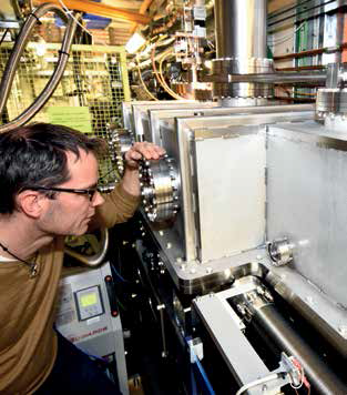
- Figure 1: PBS Thomas Sorensen on the VMXi beamline.
VMXi will be the first beamline of its kind solely dedicated to data collection directly from crystallisation experiments in situ i.e. from crystals remaining in their crystallisation media, rather than being transferred to a sample holder and exposed to X-rays under cryogenic conditions. The beamline will have facilities to store thousands of user crystallisation experiments and will feature automated transfer between sample storage and the beamline, as well as highly automated data collection and analysis. It will enable users to monitor their crystallisation experiments remotely and request X-ray analysis without the need for their direct participation in the X-ray experiment. This approach will allow for the study of crystals as they emerge and for the collection of data from all crystals, including those that are too fragile to handle and those that cannot be cryo-cooled. VMXi is due to become available to users at the end of 2016.
I14 - Hard X-ray Nanoprobe
At over 185m long I14 is a scanning probe beamline that uses X-ray fluorescence and diffraction techniques to determine the composition of a wide range of materials. It will produce X-ray beams of 30-50 nm with energies higher than 5 keV, giving better penetration depth for thick or wet samples. This will open up the possibility of studying micron scale samples in greater detail, such as biological cells that are generally on the scale of 1-5 μm. The construction of the external building was completed in February 2016 and installation and testing will continue through 2016 with first users expected in spring 2017.
I21 - Inelastic Soft X-ray Scattering
I21 is a dedicated Inelastic X-ray Scattering (IXS) facility that will produce highly monochromatised, focused and tunable X-ray beams. It is suited to investigate the electronic, magnetic and lattice dynamics of samples particularly those with magnetic and electronic interactions. The beamline will be 81 m long, with its end station and spectrometer accommodated in an external building adjacent to the Diamond ring. First light was achieved in December 2015, two months ahead of the original milestone. This year will see staged X-ray commissioning from the internal beamline towards the exit slit in spring, the sample vessel by autumn. Final X-ray commissioning in the grating and detector assembly is expected by January 2017.
B07 - Versatile Soft X-rays (VERSOX)
VERSOX will offer X-ray Photoelectron Spectroscopy (XPS) and X-ray Absorption Spectroscopy (XAS) at high pressures. The beamline will offer users the ability to carry out atmospheric chemistry, catalysis, and biological investigations with samples under native conditions such as liquid environments or gas-phase reaction conditions for catalysts. The last year has seen significant progress to B07’s construction. In 2015 the end station was commissioned at I09 and was open to users from February 2016 until March 2016. It is expected that this will enable a fast transfer of the end station when the beamline at B07 is commissioned later in the year.
I02-1 - Versatile MX (VMXm)
VMXm will perform atomic resolution structure determination from micro and nanocrystals of biological macromolecules that give rise to very weak diffraction. This is typically the case for crystals of flexible, complex biological macromolecules. The minimum X-ray beam size on VMXm will be less than 0.5 microns and will uniquely combine scanning electron microscopy with X-ray diffraction to allow nanocrystals to be sequentially aligned into the X-ray beam for data collection. VMXm is currently under construction and is scheduled for first user operations in January 2018.
DIAD: Dual Imaging and Diffraction
DIAD is based on an innovative X-ray optical concept to allow the study of in situ processes with both imaging and diffraction simultaneously, enabling the user to take measurements of a live process as it evolves. Obtaining microscopic resolution, the instrument will serve those interested in the spatial and time-evolution of the structural properties of samples that undergo a range of treatments such as mechanical, chemical or thermal. Research will cover the bio-medical field (bone and tissue), material science (composite materials, damage), chemistry (catalysis, corrosion), geological science (rock formation), biology (plant science, soil interactions) and energy (batteries, irradiated materials).

Diamond Light Source is the UK's national synchrotron science facility, located at the Harwell Science and Innovation Campus in Oxfordshire.
Diamond Light Source Ltd
Diamond House
Harwell Science & Innovation Campus
Didcot
Oxfordshire
OX11 0DE
Copyright © Diamond Light Source. Diamond Light Source® and the Diamond logo are registered trademarks of Diamond Light Source Ltd
Registered in England and Wales at Diamond House, Harwell Science and Innovation Campus, Didcot, Oxfordshire, OX11 0DE, United Kingdom. Company number: 4375679. VAT number: 287 461 957. Economic Operators Registration and Identification (EORI) number: GB287461957003.