Unraveling iron uptake and magnetosome formation in magnetospirillum gryphiswaldense
May 8, 2025
May 8, 2025
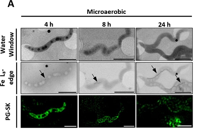
Iron plays several essential roles in bacteria, making it a crucial element for their survival and function. In magnetotactic bacteria like Magnetospirillum gryphiswaldense, iron plays a central role in the formation of magnetosomes. These peculiar bacteria possess the capability to orient themselves along the Earth's magnetic field lines, thanks to the presence of a very specific type of intracellular magnetic nanoparticles called magnetosomes. Magnetosomes are mainly composed of magnetite crystals (Fe3O4) enveloped in a lipidic membrane. Some mechanisms such as the internalisation and the transformation of iron into magnetite crystals are still poorly understood. In an article recently published in ACS Applied Materials & Interfaces, a team of researchers from Aston University investigated the formation of these magnetosomes in bacteria by finely tuning the concentration of oxygen and iron. They performed CryoSIM and CryoSXT experiments on the B24 beamline. The team were also the first to exploit the recent development of the beamline to measure X-ray absorption data at the Iron L3 edge to aid visualisation of the magnetsomes.
Magnetosome formation in magnetotactic bacteria is a complex process influenced by environmental factors such as iron concentration and oxygen levels. Prior studies provided foundational knowledge but lacked the resolution to observe these processes at the single-cell level under near-native conditions. Given the small size of magnetosomes, which can range from 30 nm to 120 nm across different species, electron microscopy is one of the most common used imaging techniques. However, this approach does not enable simultaneous tracking of intracellular iron content alongside magnetosome content to understand better how the biomineralisation process works. This research aimed to bridge that gap by employing an integrated approach combining correlative light and X-ray microscopy with other analytical techniques.
Firstly, the data obtained from these other analytical techniques suggested a potential correlation between the intracellular iron pool and magnetosome content. Specifically, increased iron availability under microaerobic conditions appeared to result in longer magnetosome chains and higher intracellular iron concentrations. To further investigate and validate this hypothesis at the single-cell level, the researchers conducted experiments at the B24 beamline at Diamond.
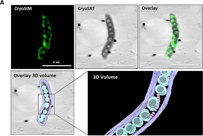
Cryo-SXT is a powerful technique used to observe the internal structure of biological samples in a near-native state. This technique uses soft-X rays to obtain three-dimensional (3D) tomograms of biological specimens with a resolution of up to 25 nm, without the need for traditional sample preparation methods that could damage cellular structures (such as drying, chemical fixation, staining). On B24, the team was able to observe internal compartments, including magnetosomes, using the preferential absorption of carbon atoms in the cell. With cryoSIM, they stained the bacteria with PG-SK, a green fluorophore that reacts with the intracellular iron. The strength of the B24 beamline is that scientists were able to analyse the same region of interest in the same samples with both CryoSIM and CryoSXT and correlate the data.
This approach provided compelling evidence of a correlation between the intracellular iron concentration and the number of magnetosomes. Another advantage of using soft X-ray microscopy at B24 is the ability to adjust the X-ray energy to the iron absorption edge. As iron atoms strongly absorb X-rays at this energy, it facilitates the observation of magnetosomes within the bacteria. By modifying the iron concentration during bacterial growth, the researchers demonstrated that these bacteria can tolerate high extracellular iron concentrations. They also identified an iron threshold beyond which increasing the extracellular iron concentration no longer leads to additional iron uptake or an increase in magnetosome production.
Understanding how magnetotactic bacteria and magnetosomes grow is essential as there is a growing interest into using magnetosomes for biotechnological and biomedical applications due to their unique properties such as narrow size distribution and biocompatibility. Building on this, the researchers plan to use the capabilities of B24 beamline for future studies that explore the use of magnetotactic bacteria as medical devices to deliver therapeutic cargo to cancer tumours. Characterising MTB and cancer cells under near-native conditions, they aim to gain critical insights into their interactions, which could enhance the development of more effective, targeted therapies for next-generation medicines.
To find out more about the B24 beamline please contact the Principal Beamline Scientist Valentina Loconte: [email protected]
Masó-Martínez, M. An Integrated Approach to Elucidate the Interplay between Iron Uptake Dynamics and Magnetosome Formation at the Single-Cell Level in Magnetospirillum gryphiswaldense ACS Appl. Mater. Interfaces 2024 DOI: 10.1021/acsami.4c15975
Image credits: this publication 10.1021/acsami.4c15975 under CC-BY 4.0 license
This article was prepared with the help of both authors: Alfred Fernandez-Castane and Marta Maso Martinez.

Diamond Light Source is the UK's national synchrotron science facility, located at the Harwell Science and Innovation Campus in Oxfordshire.
Diamond Light Source Ltd
Diamond House
Harwell Science & Innovation Campus
Didcot
Oxfordshire
OX11 0DE
Copyright © Diamond Light Source. Diamond Light Source® and the Diamond logo are registered trademarks of Diamond Light Source Ltd
Registered in England and Wales at Diamond House, Harwell Science and Innovation Campus, Didcot, Oxfordshire, OX11 0DE, United Kingdom. Company number: 4375679. VAT number: 287 461 957. Economic Operators Registration and Identification (EORI) number: GB287461957003.