How will neutron irradiation affect the superconducting magnets in fusion power plants?
Nov 30, 2022
ePSIC hosts an annual workshop on data analysis software based on the HyperSpy package. By promoting the use of open-source packages in the user community, the ePSIC team hope to encourage accessible, reliable, and shareable data analysis routines to the wider scientific community.
Nov 30, 2022
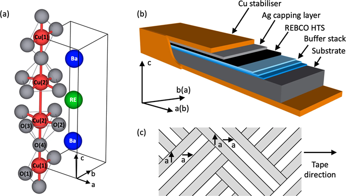
A tokamak uses a powerful magnetic field to confine plasma in a doughnut shape (torus), and is one of the concepts being developed for commercial fusion power plants. For example, the UK Atomic Energy Authority's STEP programme aims to deliver a prototype fusion energy plant by 2040. The most promising high-temperature superconducting materials suitable for making high-field magnets for small fusion tokamaks are rare-earth barium copper oxides (REBCO). However, tokamaks produce high energy neutrons and a significant flux of gamma rays, and our current understanding of how REBCO are affected by irradiation is limited. In work recently published in Communications Materials, researchers from the University of Oxford and Diamond Light Source used scanning transmission electron microscopy (STEM) at ePSIC, and High Energy Resolution Fluorescence Detected X-ray Absorption Spectroscopy (HERFD-XAS) on Diamond's I20-Scanning beamline to probe how REBCO are affected by ion irradiation. Their results challenge previous assumptions about the types of defect created, and show that annealing allows partial recovery.
Prof Susannah Speller, of the Materials Department at the University of Oxford, is the team leader of the project. She says,
REBCO are fantastic materials for making very high-temperature superconductors, which are absolutely essential for the next generation of commercial fusion reactors. But in these compact tokamaks, there's far less space for neutron shielding, and the magnets will be much closer to the plasma. That means a much higher flux of high energy neutrons penetrating the magnets. So we need to understand how REBCO will behave, and - critically - how long these superconducting materials will last in that environment. And so we've used various characterisation techniques to understand irradiation damage to the crystal structure of REBCO.
To begin with, the team used high-resolution scanning transmission electron microscopy (STEM) at ePSIC, looking at pristine, ion-irradiated materials. This technique allowed them to see, in atomic resolution, the ion columns in the material. And they found that - even in the materials that had been irradiated to the point where they stopped superconducting - the heavy ions (copper, barium and rare earth) remain in their crystallographic positions. Their results suggested that the damage occurs mainly in the oxygen sub-lattice, which is particularly difficult to see with electron microscopy. However, a complementary synchrotron X-ray technique could give them more information. X-ray Absorption Spectroscopy is a bulk technique for probing the electronic structure and the bonding environment around specific atoms, and proved to be a powerful tool for determining the nature of the radiation-induced point defects that were difficult to see with STEM.
Dr Sofia Diaz-Moreno, the Science Group Leader for Diamond's Spectroscopy Group and team member says,
Prof Speller contacted me because she had been researching a technique - High Energy Resolution Fluorescence Detection Spectroscopy - that could in principle allow us to see the REBCO structure at extremely high resolution. Specifically, the oxygen structure around the copper ions and how this environment changes after irradiation. So we designed an initial experiment for the I20-Scanning beamline, and found that it did indeed allow us to see the differences between pristine and irradiated samples. The differences between the samples are subtle, but they are enhanced by using the emission spectrometer available in the beamline.
Prof Rebecca Nicholls of Oxford University performed the density functional theory (DFT) modelling that was key to understanding the results. Prof Speller continued:
HERFD-XAS shows us the changes in the absorption edge of copper, however, in REBCO, the absorption edge of copper is very complex, with different copper sites within the crystal structure contributing peaks and features that overlap. So to interpret the results, we did some specialist DFT modelling, simulating what we'd expect to see from the pristine material, and then simulating defects in the crystal structure and the effect those would have on the spectrum.
Prior to this research, the hypothesis was that irradiation damage would be most likely to remove an oxygen from copper chains within the crystal structure. However, one of the most interesting conclusions from this research is that it creates a lot of defects in the copper-oxygen planes. Since the planes are responsible for superconductivity, it's not surprising that the superconductivity is strongly affected.
These particular experiments involved REBCO materials irradiated with He+ ions. Ion irradiation is often used as a proxy for neutron radiation, as it doesn't make materials radioactive. However, we don't yet know whether it produces the same defects in REBCO as neutron irradiation. To find out, Prof Speller's team is now working with REBCO samples irradiated in the TRIGA fission reactor in Vienna. Most labs can't handle these radioactive samples, but Diamond has opened a new Active Materials Laboratory (AML) to allow users to work with them. Prof Speller explained:
At the AML, we were able to do some initial etching of the samples to remove unwanted layers and then put them into containment to transport them to the beamline, and then we could take them back into the AML, perform an anneal to test defect recovery and take them back to the beamline to examine the results. We were the first users at the AML, and we can only investigate these neutron-irradiated materials because we have access to this facility.
Dr Mohsen Danaie is a senior electron microscopy scientist at ePSIC. He says,
This research shows the value of using complementary techniques. Being housed next to Diamond puts ePSIC in a pretty rare position, and we're always exploring how to achieve new science using a complementary electron and synchrotron radiation approach. Another key aspect of this work was using the open-source Atomap software to analyse the microscopy data, meaning the whole process from data to conclusion is transparent.
The ultimate goal of this ongoing research is to give engineers the data they need to design commercial tokamaks, such as the likely lifespan of a superconducting magnet, the amount of neutron shielding required, and any potential benefits from including an anneal in scheduled maintenance operations. A thorough understanding of those parameters brings commercial fusion power plants a step closer to reality.
To find out more about the I20 beamline or discuss potential applications, please contact Fred Mosselmans: [email protected]. For ePSIC, contact Principal Electron Microscopist Chris Allen: [email protected].
Nicholls RJ et al. Understanding irradiation damage in high-temperature superconductors for fusion reactors using high resolution X-ray absorption spectroscopy. Communications Materials 3.1: 1-14 (2022). DOI:10.1038/s43246-022-00272-0.

Diamond Light Source is the UK's national synchrotron science facility, located at the Harwell Science and Innovation Campus in Oxfordshire.
Diamond Light Source Ltd
Diamond House
Harwell Science & Innovation Campus
Didcot
Oxfordshire
OX11 0DE
Copyright © Diamond Light Source. Diamond Light Source® and the Diamond logo are registered trademarks of Diamond Light Source Ltd
Registered in England and Wales at Diamond House, Harwell Science and Innovation Campus, Didcot, Oxfordshire, OX11 0DE, United Kingdom. Company number: 4375679. VAT number: 287 461 957. Economic Operators Registration and Identification (EORI) number: GB287461957003.