Materials beamline helps take a closer look at our eyes
Oct 13, 2022
Oct 13, 2022
If you give a scientist a new piece of material and ask them to tell you about its properties, it won’t be long before they are pulling and stretching it. The technical term is “putting the material under strain” and doing this can reveal a lot about what’s inside. This conjures up images of springs, modern polymers and alloys being pulled apart by a metal vice, but you wouldn’t necessarily think about the human eye. You should.

The eye is a feat of engineering from nature that we have yet to replicate. Understanding it can give us inspiration for new technologies as well as understanding debilitating diseases. Scientists have been applying techniques from material science to help us understand the physiology of the eye. Decades ago, Professor Keith Meek, Cardiff University, pioneered the use of synchrotron X-ray scattering for the study of the cornea to better understand the structure and function of the tissue.[1]
This approach led to an understanding of how the cornea can be transparent, despite being made of the same fundamental components as skin and other tissues in our bodies, which are notably not transparent. The collagen in corneas has a specific crystalline structure which allows visible light to pass through.
 The cornea isn’t just a transparent window, however. As the main lens of the eye, it has to maintain its shape carefully, which isn’t easy as the pressure in our eyes fluctuates. There are many intricate structures in the cornea, and more recently scientists have been using synchrotron X-rays to elucidate their function and clinical relevance. This has led to a push in recent years to find ways to make it possible to gather data from biological samples in as close to natural conditions as possible.
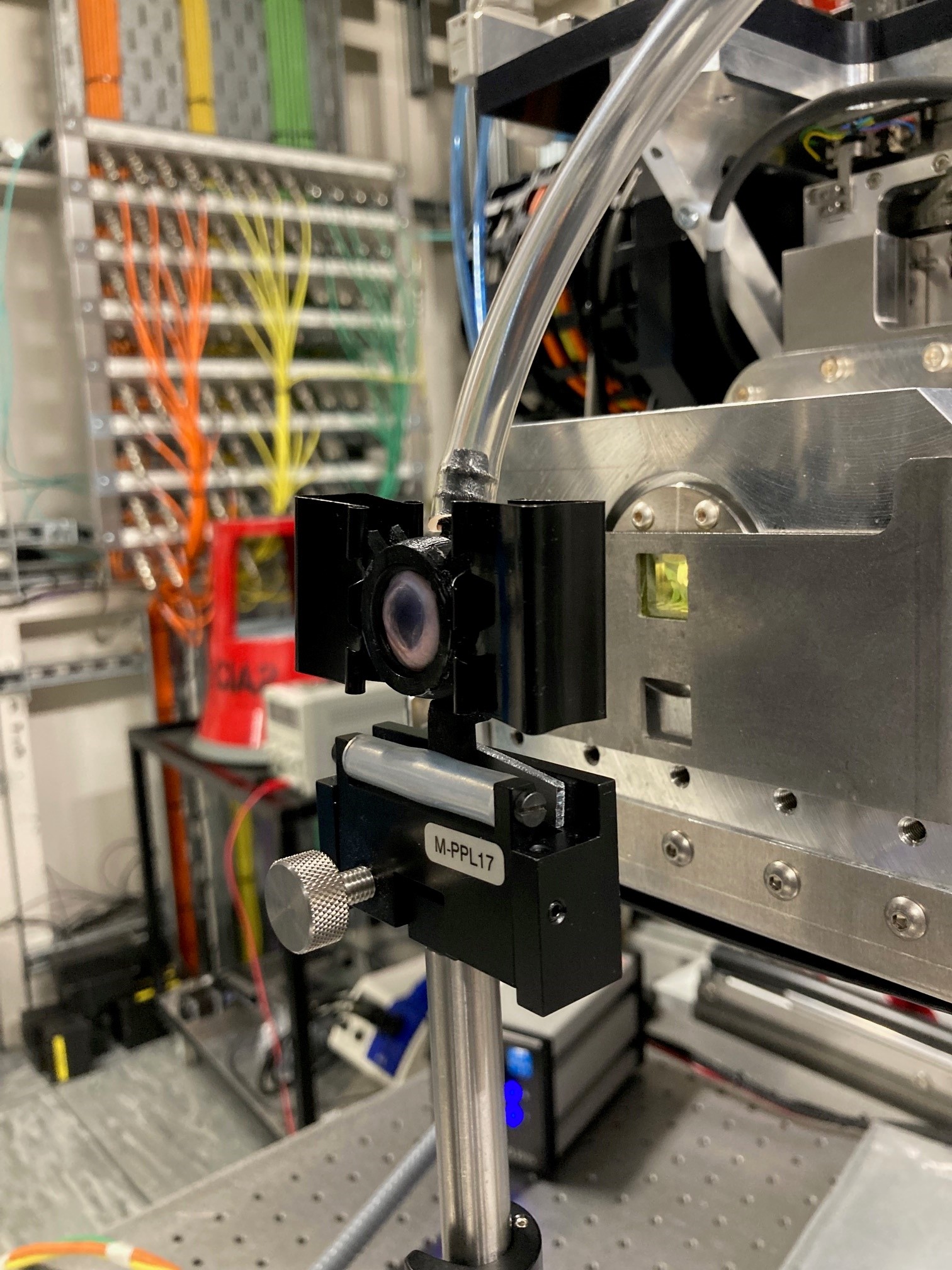
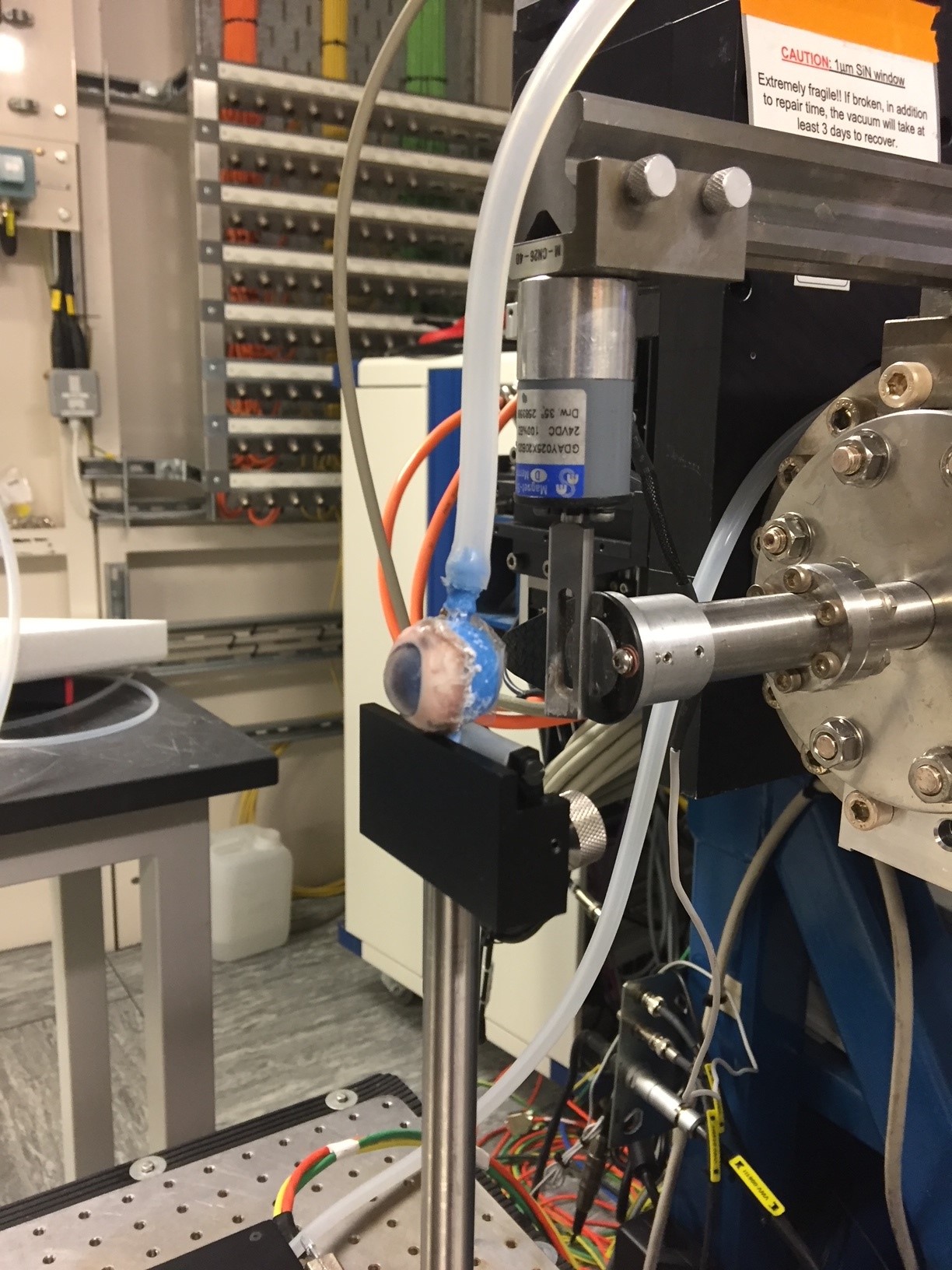
Dr James Bell from Cardiff University has made it his mission to create the most realistic environment possible to study the structural and biomechanical properties of the eye in a synchrotron beam. Over many iterations, Bell and his research team developed a rig that could inflate the cornea, adding strain in three dimensions which could then be measured using synchrotron light.
There are many different beamlines that could be used to image the eye, but for looking at the hierarchical structure of the cornea in physiological conditions, the unique combination of both Small Angle X-ray Scattering (SAXS) and Wide Angle X-ray Scattering (WAXS) at Diamond’s I22 beamline was an obvious choice. Dr Tim Snow who is a data analyst at I22 remembers vividly walking to the beamline one Wednesday morning and seeing an eye mounted to a plastic rod and gently inflating. Not your average day at the office!
 The combination of an accurate and biologically relevant rig to inflate the cornea and SAXS imaging proved to be a winning combination. The research team has embarked on a project investigating keratoconus. With this disease, the cornea gradually deforms into a cone and if left untreated, can require a corneal transplant which are, unfortunately, in very high demand.
However, there is a treatment for keratoconus if it is caught in time. You can crosslink the proteins in the eye making it stiffer and preventing further change in shape. The problem is that the treatment, although effective in most cases, is still not well understood and there is no established code of best practice. Studies at I22 will continue to provide structural data to improve the accuracy of computational models aimed at predicting and improving treatment outcomes.
The work by James and his team including Sally Hayes, Keith Meek, Olga Shebanova (from I22) and Siân Morgan, will continue as part of a five-year MRC grant to understand the cornea and to develop new therapies.
[1] a) K. M. Meek, G. F. Elliott, Z. Sayers, S. B. Whitburn and M. H. J. Koch, Journal of Molecular Biology 1981, 149, 477-488; b) K. M. Meek, Biophysical Reviews 2009, 1, 83-93.
[2] a) J. Bell, S. R. Morgan, E. Koudouna, S. Hayes and K. M. Meek, Investigative Ophthalmology & Visual Science 2022, 63, 2392 – A0195-2392 – A0195; b) J. S. Bell, S. Hayes, C. Whitford, J. Sanchez-Weatherby, O. Shebanova, N. J. Terrill, T. Sorensen, A. Elsheikh and K. M. Meek, Acta Biomaterialia 2022, 8.

Diamond Light Source is the UK's national synchrotron science facility, located at the Harwell Science and Innovation Campus in Oxfordshire.
Diamond Light Source Ltd
Diamond House
Harwell Science & Innovation Campus
Didcot
Oxfordshire
OX11 0DE
Copyright © Diamond Light Source. Diamond Light Source® and the Diamond logo are registered trademarks of Diamond Light Source Ltd
Registered in England and Wales at Diamond House, Harwell Science and Innovation Campus, Didcot, Oxfordshire, OX11 0DE, United Kingdom. Company number: 4375679. VAT number: 287 461 957. Economic Operators Registration and Identification (EORI) number: GB287461957003.