Investigating X-ray damage to small molecular crystals
Sep 10, 2021
Sep 10, 2021
X-ray imaging and characterisation techniques are such valuable tools for discovery that we see their use expanding throughout the scientific community. While laboratory X-ray sources are achieving small beam sizes and high photon flux density comparable to today's synchrotron sources, new fourth-generation synchrotrons - with higher brightness X-ray beams - are coming online. In addition, an increasing number of X-ray Free Electron Lasers (XFELs) offer ultra-short (fs), coherent pulses of X-ray radiation of superior brilliance. As the power of our X-ray probes increases, so does the importance of understanding the effects of radiation damage on samples. While significant advances have been made in understanding the radiation damage processes in macromolecular crystallography, the effects of X-ray exposure on small molecular crystals have been largely overlooked. In work recently published in the Journal of Physical Chemistry, a team of researchers conducted systematic studies of X-ray-matter interactions for two industrially important catalysts exposed to radiation in X-ray diffraction (XRD) and X-ray photoelectron spectroscopy (XPS) experiments. Their combined techniques offer important insights into the X-ray induced effects in transition metal catalysts and their intrinsic stabilities, and can be applied to other small molecular systems of scientific or industrial relevance.
![Schematic showing the molecular structure of [M(COD)Cl]2 and the different processes occurring upon X-ray irradiation during diffraction and photoelectron spectroscopy experiments.](/dam/jcr:0eda168b-8ff0-4685-b4ca-f5362a888fae/I111.png)
Researchers bring all kinds of samples to Diamond and other synchrotrons, to examine and analyse their physical structure and chemical make-up. Powerful X-ray beams are invaluable probes of matter and have played a role in many significant scientific and technological developments. However, we know that X-rays can cause radiation damage to samples, and this issue becomes more important as X-ray sources get stronger. There's also a trend towards more dynamic experiments that watch effects unfold in the X-ray beam, with longer exposures potentially leading to more sample damage.
Large-scale studies have expanded our understanding of radiation damage processes in macromolecular crystallography (MX). This, in turn, has allowed the development of mitigation strategies, including the incorporation of molecular radioprotectants and scavengers, beam attenuation and sample cryocooling. However, to date, there have been few corresponding systematic studies of radiation damage in small molecular systems or framework materials, ceramics and alloys.
In this research, scientists from University College London, Imperial College London, the University of Oxford, the MRC Laboratory of Molecular Biology and Diamond conducted systematic studies of X-ray-matter interactions on two industrially important catalysts, [Ir(COD)Cl]2 and [Rh(COD)Cl]2, exposed to radiation in X-ray diffraction (XRD) and X-ray photoelectron spectroscopy (XPS) experiments.
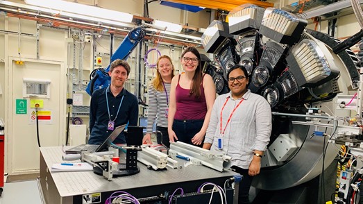
To begin with, they carried out single crystal XRD studies of both catalysts on the I19 beamline. This gave them the starting structures and a starting point for the powder XRD diffraction experiments. Then, moving to beamline I11, they carried out iterative experiments, with crystals exposed for two hours and diffraction patterns collected every couple of seconds. This allowed them to see how the crystal structure changed over time.
Senior author Dr Anna Regoutz explains:
Our experiments on I11 were crucial to this project. I11 has a high photon flux, and a very efficient detector system, so you can collect results quickly. Usually, radiation damage causes a crystal to become less ordered and crystalline, which means the quality of the data collected is often lower. Using I11 allowed us to continue to collect high-quality data as the materials degraded. And in two hours we get 500 datasets, which allows us to follow changes on a relevant timescale. We can see the trends continuously. If we tried to get the same results from single crystal XRD, it would take considerably longer.
The team combined their synchrotron studies with laboratory-based X-ray photoelectron spectroscopy (XPS) at Imperial College London. XRD provides an average picture of the changes in the crystal structure, bond lengths and angles. Using XPS adds detail about the local changes in the chemical environment and electronic structure.
Lead author Nathalie Fernando is a PhD student at University College London. She says:
We were able to correlate the changes in the XRD and XPS experiments to show the relationship between the structural changes and the changes in the chemical environment. It's vital to have this information, because when researchers conduct X-ray experiments they have to be able to account for any radiation damage in the sample to get reliable results.
The results showed radiation-induced changes, in both catalysts, to the physical structure and local chemical environment, which manifested in changes to the electronic structure. They also provide further evidence that established mitigation strategies are effective. However, the results highlight the importance of choosing the best experimental setup and parameters, as these vary between techniques and beamlines and can substantially affect radiation dose and damage.
This work also offers important insights into the intrinsic stability of transition metal catalysts. The subjects chosen are members of the [M(COD)Cl]2 family of prototypical catalysts. The two samples have the same ligand structure and comparable valence electronic structure, but different metal centres. They also have different absorption coefficients at the photon energies used, leading to substantial differences in the absorbed X-ray dose and the subsequent damage.
Dr Regoutz concludes:
For most experiments, researchers will want to reduce radiation damage, and our results will help with that. However, because we used two related catalysts, we've learned something about the intrinsic stabilities of these systems. By doing a systematic study, we can learn how different molecular systems react to radiation, which might tell us something about their final stability in an application. And from there, we can think about tuning materials to be stable for a specific application.
To find out more about the I11 beamline or discuss potential applications, please contact Principal Beamline Scientist (PBS) Stephen Thompson: [email protected]. The PBS for I19 is Dave Allan: [email protected].
Fernando NK et al. Structural and Electronic Effects of X-ray Irradiation on Prototypical [M (COD) Cl] 2 Catalysts. The Journal of Physical Chemistry A 125, 34, 7473–7488 (2021). DOI: 10.1021/acs.jpca.1c05759.
(Also available via ChemRxiv.)

Diamond Light Source is the UK's national synchrotron science facility, located at the Harwell Science and Innovation Campus in Oxfordshire.
Diamond Light Source Ltd
Diamond House
Harwell Science & Innovation Campus
Didcot
Oxfordshire
OX11 0DE
Copyright © Diamond Light Source. Diamond Light Source® and the Diamond logo are registered trademarks of Diamond Light Source Ltd
Registered in England and Wales at Diamond House, Harwell Science and Innovation Campus, Didcot, Oxfordshire, OX11 0DE, United Kingdom. Company number: 4375679. VAT number: 287 461 957. Economic Operators Registration and Identification (EORI) number: GB287461957003.