Advanced X-ray imaging using sandpaper
Mar 18, 2015
Mar 18, 2015
Scientists working on Diamond Light Source’s dedicated test beamline, B16, have developed a new X-ray dark-field imaging technique allowing images of materials in a hitherto unforeseen level of simplicity. The results of their research have been recently published in Physical Review Letters.
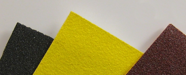
The abrasive paper used in the experimental setup on B16
Conventional X-ray imaging, known as absorption imaging, takes advantage of the fact that a material has a distinctly different density to its surroundings. An image of a human skeleton is possible as the bones absorb the X-rays passing through the body far more than the tissue around them. The X-ray dark-field imaging in contrast employs the different scattering power of different parts of a sample to provide additional and complementary information even though they may have similar absorption contrast.
Using everyday materials, such as sandpaper from a DIY store, the methodology takes advantage of the speckle technique commonly used in laser interferometry, and could lead to easy to use and cheaper imaging devices.
"There are no special optics involved – we only need a sheet of abrasive paper to generate the speckle pattern and an X-ray camera." says Dr Hongchang Wang, Senior Optics Scientist at Diamond and lead author of two recent papers on the technique. "It’s a simple experimental setup, and the abrasive paper is cheap, robust and commercially available."
However, producing images of soft tissues is more complex, as there is the range of densities required to produce an imaging using the absorption method. Phase contrast imaging allows the imaging of structures of similar levels of transparency by highlighting small differences in the refractive indexes of the materials within the structure.
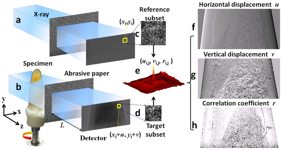
Speckle image (a) without the specimen and (b) with the specimen and the phase object (abrasive paper), (c) reference and (d) target subset speckle image, (e) example of correlation coefficient map for the subset, (f) horizontal and (g) vertical displacement and (h) correlation coefficient image
Wang H. et al. X‐ray phase contrast tomography by tracking near field speckle. Sci Reports (2015) 10.1038/srep08762
In order to reveal the internal structure of soft tissues, the team has also extended the speckle based phase contrast imaging from 2D radiography to 3D tomography, and while the initial work was carried out on B16, Diamond scientists teamed up with scientists from PETRA III (Hamburg) to perform further experiments on PETRA’s P05 beamline to produce the first 3D image of a human artery by using speckle based technique. As described in their publication in Scientific Reports, significant enhanced contrast has been observed in the phase contrast computed tomography (CT) compared with the conventional absorption contrast CT. The simplicity of the experimental arrangement and speed of measurement gives this new imaging method a distinct advantage over existing X-ray imaging methods, and as such, make it an attractive technique for in vivo imaging of soft-tissue biological systems.
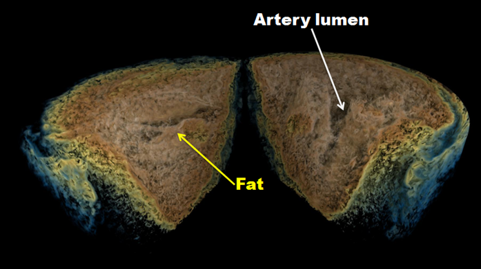
3D rendering of human artery: distinctive contrast can be observed between the artery lumen and fat
"This represents a significant step forward, and has been able to progress due to the flexible capabilities of B16." explains Dr Kawal Sawhney, the Principal Beamline Scientist on B16 who is also a co-author on two papers on the new imaging technique. "Ultimately, we hope to show how to develop a quick and simple technique that will help people easily image more complex samples."
The research team plan to continue to develop the technique on the versatile platform that the B16 beamline offers, hoping to make advances that allow it to be performed with X-rays from table top devices. The technique may offer solutions to imaging problems in medicine, materials science and security.
For further information on the B16 test beamline or to discuss potential applications, please contact Principal Beamline Scientist Dr Kawal Sawhney: [email protected]
Wang H. Kashyap Y. & Sawhney K. Hard X-ray directional dark-field imaging using speckle scanning technique. Phys Rev Lett. (2015) DOI: 10.1103/PhysRevLett.114.103901
Wang H. Berujon S. Herzen J. Atwood R. Laundy D. Hipp A. & Sawhney K. X‐ray phase contrast tomography by tracking near field speckle. Sci Reports (2015) DOI: 10.1038/srep08762

Diamond Light Source is the UK's national synchrotron science facility, located at the Harwell Science and Innovation Campus in Oxfordshire.
Diamond Light Source Ltd
Diamond House
Harwell Science & Innovation Campus
Didcot
Oxfordshire
OX11 0DE
Copyright © Diamond Light Source. Diamond Light Source® and the Diamond logo are registered trademarks of Diamond Light Source Ltd
Registered in England and Wales at Diamond House, Harwell Science and Innovation Campus, Didcot, Oxfordshire, OX11 0DE, United Kingdom. Company number: 4375679. VAT number: 287 461 957. Economic Operators Registration and Identification (EORI) number: GB287461957003.